
Classification of Tissues MCQs for GPAT and Pharmacist Exams
Prepare for NIPER, AIIMS Pharmacist, Railway, SSC, ESIC and State Pharmacist exams with quick revision notes and MCQs on Classification of Tissues MCQs for GPAT and Pharmacist Exams structure, location and functions of epithelial, connective, muscular and nervous tissues.
Dr. Alok Singh
7/11/20265 min read
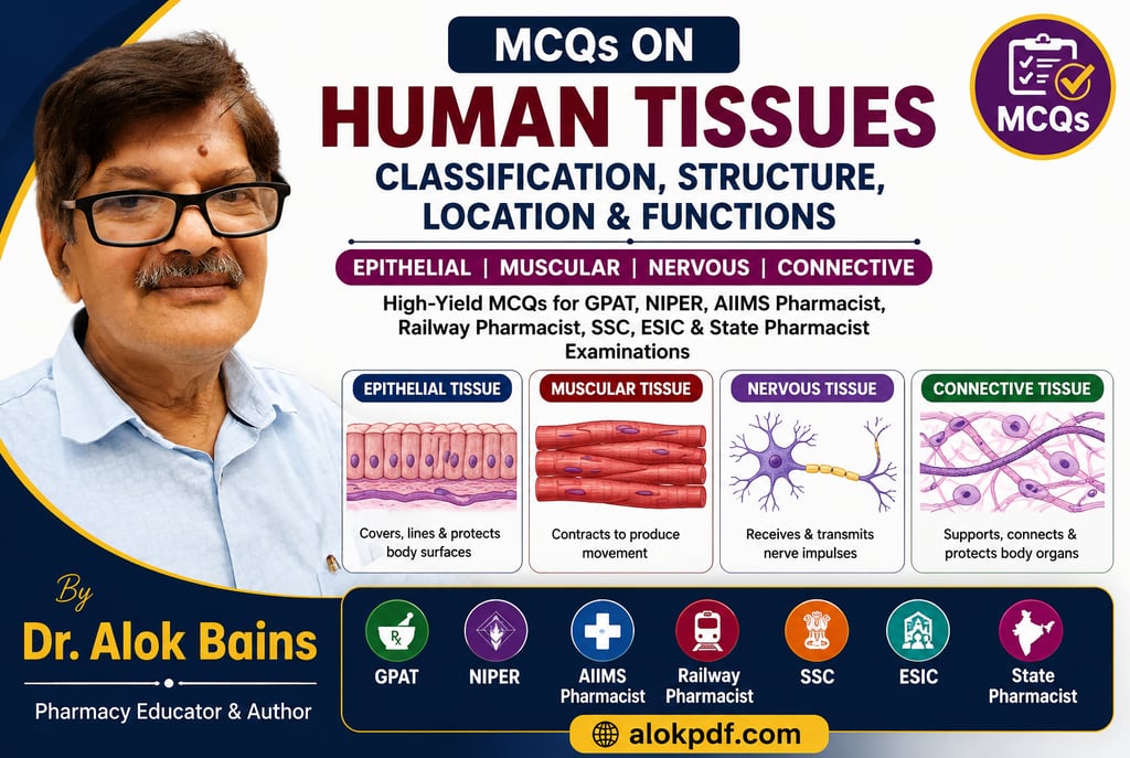
MCQs for GPAT, NIPER, AIIMS Pharmacist, Railway Pharmacist, SSC, ESIC, and State Pharmacist examinations: Human Tissues.
Quick Revision Notes: Classification of Tissues
What is tissue?
A tissue is a group of similar cells along with their intercellular substance that work together to perform a specific function.
The human body contains four basic types of tissues:
Epithelial Tissue
Connective Tissue
Muscular Tissue
Nervous Tissue
A simple way to remember them is:
E-C-M-N → "Every Cell Must Network"
1. Epithelial Tissue
Epithelial tissue forms the covering of body surfaces, lines body cavities and ducts, and forms glands.
Important Structural Features
Cells are closely packed.
Very little intercellular matrix is present.
Rest on a basement membrane.
Usually avascular (lacks blood vessels).
Shows rapid regeneration.
Classification of Epithelial Tissue
A. Simple Epithelium (single layer)
Type Structure Location Function
Simple squamous Flat cells Alveoli, Bowman's capsule, blood vessels Diffusion & filtration
Simple cuboidal Cube-shaped cells Kidney tubules, thyroid gland Secretion & absorption
Simple columnar Tall cells Stomach & intestine Absorption & secretion
Ciliated columnar Columnar cells with cilia Fallopian tubes, respiratory tract Movement of mucus/ovum
B. Stratified Epithelium (many layers)
Type Location Function
Stratified squamous Skin, mouth, esophagus Protection from abrasion
Transitional epithelium Urinary bladder & ureter Stretching and distension
High-Yield Points
Thinnest epithelium: Simple squamous.
Stretchable epithelium: Transitional epithelium.
Fastest regenerating tissue: Epithelium.
Cilia move mucus, whereas microvilli increase absorption.
2. Connective Tissue
Connective tissue supports, binds, protects, and connects different tissues and organs.
Important Structural Features
Cells are widely separated.
Contains abundant extracellular matrix.
Usually well supplied with blood vessels.
Matrix may be fluid, gel-like, or solid.
Classification of Connective Tissue
A. Connective Tissue Proper
Type Location Function
Areolar tissue Under skin & around organs Packing & support
Adipose tissue Beneath skin, around kidneys Fat storage & insulation
Reticular tissue Spleen, lymph nodes Forms supporting framework
B. Dense Connective Tissue
Type Function
Tendon Connects muscle to bone
Ligament Connects bone to bone
C. Supporting Connective Tissue
Type Location Function
Cartilage Nose, ear, trachea Flexible support
Bone Skeleton Support and protection
D. Fluid Connective Tissue
Type Function
Blood Transport of gases and nutrients
Lymph Immunity and drainage of tissue fluid
Types of Cartilage
Type Location
Hyaline cartilage Trachea, nose, costal cartilage
Elastic cartilage External ear and epiglottis
Fibrocartilage Intervertebral discs and pubic symphysis
High-Yield Points
Most abundant tissue in the body: Connective tissue.
Cartilage is avascular; therefore, it heals slowly.
Most abundant cartilage: Hyaline cartilage.
Strongest connective tissue: Bone.
3. Muscular Tissue
Muscular tissue consists of specialised cells capable of contraction and relaxation, producing movement.
Types of Muscular Tissue
Type Structure Control Location Function
Skeletal muscle Striated, long, multinucleated Voluntary Attached to bones Body movements
Smooth muscle Non-striated, spindle-shaped Involuntary Intestine, blood vessels, uterus Peristalsis & organ movement
Cardiac muscle Striated, branched, central nucleus Involuntary HeartPumping blood
High-Yield Points
Largest muscle tissue: Skeletal muscle.
Intercalated discs are present only in cardiac muscle.
Peristalsis occurs due to smooth muscle contraction.
Cardiac muscle contracts rhythmically throughout life.
Easy Memory Trick
S-S-C = Skeleton, Stomach, Cardiac
Skeleton → Skeletal muscle
Stomach → Smooth muscle
Cardiac → Heart muscle
4. Nervous Tissue
Nervous tissue receives stimuli and transmits impulses throughout the body.
Components of Nervous Tissue
A. Neurons
Functional units of the nervous system.
Parts of a Neuron
Part Function
Dendrites Receive impulses
Cell body Metabolic center
Axon Conducts impulses away from cell body
Myelin sheath Insulates axon and increases speed of conduction
Nodes of Ranvier Enable saltatory conduction
B. Neuroglial Cells
Supporting cells of the nervous system.
Cell Function
Schwann cells Form myelin in PNS
Oligodendrocytes Form myelin in CNS
Astrocytes Support and nourish neurons
Microglia Phagocytic cells of CNS
Ependymal cells Line ventricles and produce CSF
Imp. Points
Functional unit of the nervous system: Neurone.
Dendrites receive impulses; axons transmit impulses.
The myelin sheath increases conduction velocity.
Nodes of Ranvier help in saltatory conduction.
Nervous tissue has very poor regenerative capacity.
Quick Comparison of the Four Tissues
Feature Epithelial Connective Muscular Nervous
Main function Covering & secretion Support & binding Movement Communication
Cell arrangement Closely packed Widely separated Elongated fibers Neurons with processes
Matrix Very little Abundant Minimal Minimal
Blood supply Usually absent Usually rich Rich Rich
Regeneration Excellent Moderate Limited Poor
Last-Minute Revision Pearls
Simple squamous: Diffusion
Transitional epithelium: Stretching
Tendon: Muscle to bone
Ligament: Bone to bone
Smooth muscle: Peristalsis
Cardiac muscle: Intercalated discs
Schwann cell: PNS myelin
Oligodendrocyte: CNS myelin
Dendrites receive; axons send
Connective tissue is the most abundant tissue in the body
Histology MCQs for pharmacy students
1. Which tissue type is characterised by the presence of very little intercellular matrix and closely packed cells?
A. Connective tissue
B. Nervous tissue
C. Epithelial tissue
D. Muscular tissue
Answer: C
Explanation: Epithelial tissues consist of tightly packed cells with minimal extracellular matrix.
2. Which of the following epithelial tissues is specialized for diffusion and filtration?
A. Stratified squamous epithelium
B. Cuboidal epithelium
C. Simple squamous epithelium
D. Transitional epithelium
Answer: C
Explanation: Thin flattened cells facilitate rapid diffusion in alveoli and Bowman's capsule.
3. The epithelium lining the urinary bladder is:
A. Stratified cuboidal
B. Transitional epithelium
C. Simple columnar
D. Pseudostratified columnar
Answer: B
Explanation: Transitional epithelium permits stretching during bladder filling.
4. Cilia are commonly present on:
A. Simple squamous epithelium of alveoli
B. Stratified squamous epithelium of skin
C. Pseudostratified columnar epithelium of trachea
D. Transitional epithelium of ureter
Answer: C
Explanation: Cilia help move mucus and trapped particles toward the pharynx.
5. Which epithelial tissue provides maximum protection against mechanical abrasion?
A. Simple cuboidal epithelium
B. Simple columnar epithelium
C. Stratified squamous epithelium
D. Simple squamous epithelium
Answer: C
6. Microvilli are most abundant in:
A. Skin epidermis
B. Kidney tubules and intestine
C. Urinary bladder
D. Cornea
Answer: B
Explanation: Microvilli increase absorptive surface area.
7. Which statement regarding connective tissue is correct?
A. Cells are tightly packed with no matrix.
B. It contains abundant extracellular matrix.
C. It is always vascular.
D. It is incapable of repair.
Answer: B
8. Cartilage differs from bone because cartilage:
A. Is highly vascular
B. Contains osteocytes
C. Is avascular
D. Contains Haversian systems
Answer: C
9. Which connective tissue connects muscle to bone?
A. Ligament
B. Tendon
C. Cartilage
D. Areolar tissue
Answer: B
10. Which connective tissue connects bone to bone?
A. Tendon
B. Reticular tissue
C. Ligament
D. Elastic cartilage
Answer: C
11. Which type of cartilage is found in the intervertebral discs?
A. Hyaline cartilage
B. Elastic cartilage
C. Fibrocartilage
D. Calcified cartilage
Answer: C
12. Elastic cartilage is found in:
A. Trachea
B. Costal cartilage
C. External ear pinna
D. Articular surfaces of bones
Answer: C
13. Which of the following tissues has the greatest regenerative capacity?
A. Cardiac muscle
B. Nervous tissue
C. Skeletal muscle
D. Epithelial tissue
Answer: D
14. Which muscle type possesses intercalated discs?
A. Skeletal muscle
B. Smooth muscle
C. Cardiac muscle
D. Visceral muscle
Answer: C
15. The muscle responsible for peristalsis in the gastrointestinal tract is:
A. Skeletal muscle
B. Cardiac muscle
C. Smooth muscle
D. Striated voluntary muscle
Answer: C
16. Which feature distinguishes cardiac muscle from skeletal muscle?
A. Presence of striations
B. Presence of multiple nuclei
C. Branching fibers and intercalated discs
D. Voluntary control
Answer: C
17. Skeletal muscle fibres are typically
A. Non-striated and involuntary
B. Striated and multinucleated
C. Branched and uninucleated
D. Spindle-shaped and involuntary
Answer: B
18. Which muscle type can continue rhythmic contraction without nervous stimulation?
A. Skeletal muscle
B. Smooth muscle
C. Cardiac muscle
D. All of these
Answer: C
19. The functional unit of nervous tissue is:
A. Neuroglia
B. Axon
C. Dendrite
D. Neuron
Answer: D
20. Which part of the neuron receives impulses from adjacent neurons?
A. Axon
B. Myelin sheath
C. Dendrites
D. Node of Ranvier
Answer: C
21. Myelin sheath in the peripheral nervous system is formed by:
A. Astrocytes
B. Oligodendrocytes
C. Schwann cells
D. Microglia
Answer: C
22. Saltatory conduction occurs because action potentials jump between:
A. Dendrites
B. Axon hillocks
C. Nodes of Ranvier
D. Synaptic clefts
Answer: C
23. Which neuroglial cells perform phagocytic functions in the CNS?
A. Astrocytes
B. Oligodendrocytes
C. Microglia
D. Ependymal cells
Answer: C
24. Which tissue has the least regenerative ability in adults?
A. Bone tissue
B. Connective tissue
C. Nervous tissue
D. Epithelium
Answer: C
25. Match the following:
TissueFunctionP. Simple squamous1. StretchingQ. Transitional2. Rapid diffusionR. Cardiac muscle3. Pumping bloodS. Tendon4. Muscle to bone attachment
Choose the correct option:
A. P-2, Q-1, R-3, S-4
B. P-1, Q-2, R-4, S-3
C. P-2, Q-3, R-1, S-4
D. P-4, Q-2, R-3, S-1
Answer: A
26. Which tissue type forms the stroma or supporting framework of organs?
A. Epithelial tissue
B. Nervous tissue
C. Reticular connective tissue
D. Skeletal muscle
Answer: C
27. The basement membrane is located:
A. Between connective tissue and muscle
B. Between epithelium and underlying connective tissue
C. Between neuron and glial cells
D. Between tendon and muscle
Answer: B
28. Which of the following is an example of dense regular connective tissue?
A. Dermis
B. Tendon
C. Areolar tissue
D. Adipose tissue
Answer: B
29. Which statement regarding smooth muscle is FALSE?
A. It is involuntary.
B. Cells are spindle-shaped.
C. Fibres are multinucleated.
D. It is present in blood vessel walls.
Answer: C
30. A biopsy specimen showing branching striated fibres with central nuclei most likely belongs to:
A. Skeletal muscle
B. Smooth muscle
C. Cardiac muscle
D. Tendon
Answer: C
One-Liners for Competitive Exams
Most abundant tissue in the body: Connective tissue
Most abundant cartilage: Hyaline cartilage
Fastest regenerating tissue: Epithelium
Least regenerative tissue: Nervous tissue
Muscle with intercalated discs: Cardiac muscle
Neuron myelinating cell in PNS: Schwann cell
Neuron myelinating cell in CNS: Oligodendrocyte
Stretchable epithelium: Transitional epithelium
Gas exchange epithelium: Simple squamous epithelium
Peristalsis muscle: Smooth muscle
Dr. Alok Singh