
Genetic Code and DNA Repair MCQs
Practice challenging MCQs on Genetic Code and DNA Repair MCQs and DNA repair, protein synthesis inhibitors, DNA damage, repair mechanisms, and related disorders for GPAT, NIPER, AIIMS, and pharmacist exams.
Dr. Alok Singh
7/24/202614 min read
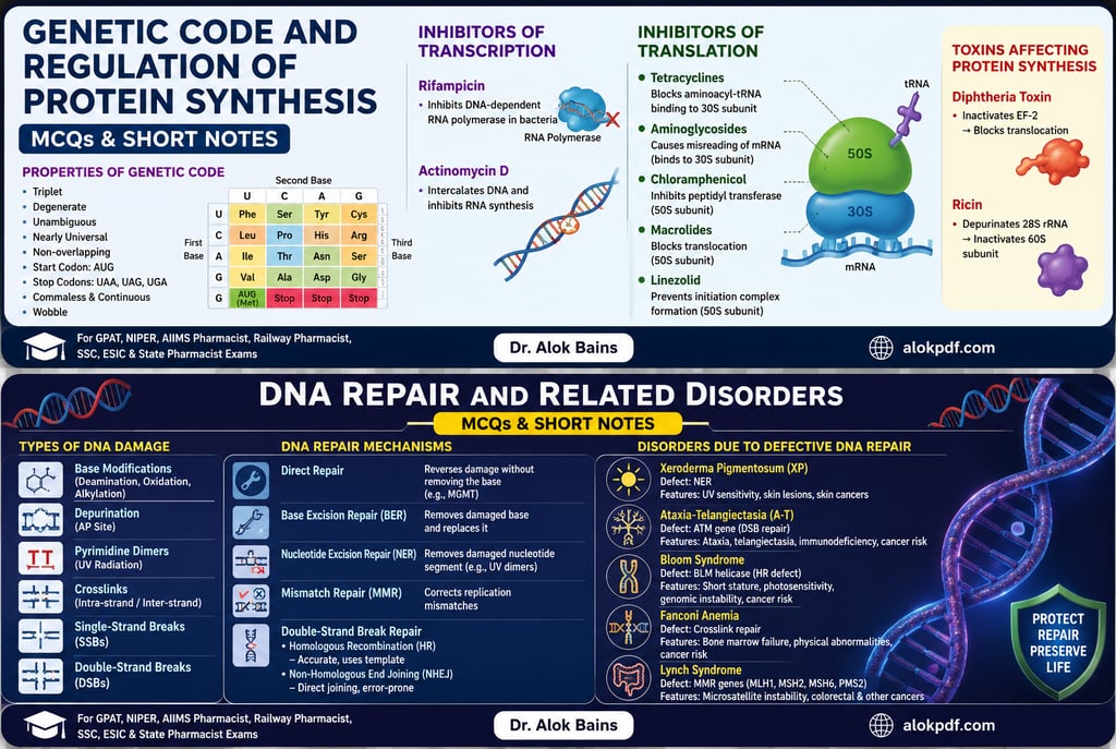
MCQs: Genetic Code, Regulation of Protein Synthesis, DNA Damage and Repair
Quick Revision Notes: Genetic Code, Protein Synthesis, DNA Repair & Related Disorders
1. Genetic Code
The genetic code is the set of rules by which the nucleotide sequence of mRNA is translated into the amino acid sequence of a protein.
Key Properties of the Genetic Code
Triplet code: Each codon contains three nucleotides and specifies one amino acid or a stop signal.
Degenerate: Most amino acids are encoded by more than one codon.
Example: Leucine, serine, and arginine each have six codons.
Unambiguous: Each codon specifies only one amino acid.
Nearly universal: The same codons generally specify the same amino acids in almost all organisms.
Exception: Some mitochondrial and other genetic codes differ slightly.
Non-overlapping: A nucleotide is normally read as part of only one codon within a given reading frame.
Commaless/continuous: Codons are read continuously without gaps or punctuation.
Start codon: AUG initiates translation and codes for methionine (formylmethionine in bacteria).
Stop codons: UAA, UAG, and UGA signal termination of translation.
Wobble: Flexibility at the third base of the codon allows one tRNA to recognize more than one codon.
Easy Memory Tip
"AUG Starts; UAA, UAG, UGA Stop."
2. Regulation and Inhibition of Protein Synthesis
Protein synthesis occurs in two major stages:
DNA → Transcription → mRNA → Translation → Protein
Transcription: Formation of RNA from a DNA template.
Translation: Formation of protein using the mRNA sequence at the ribosome.
Important Inhibitors of Transcription
Rifampicin (Rifampin)
Acts mainly on bacteria.
Binds to bacterial DNA-dependent RNA polymerase.
Inhibits transcription by preventing RNA synthesis.
Exam point:
Rifampicin → RNA polymerase → Transcription blocked
Actinomycin D (Dactinomycin)
Intercalates between DNA base pairs.
Prevents movement of RNA polymerase along DNA.
Mainly inhibits transcription.
Used clinically as an antineoplastic drug.
Important Inhibitors of Translation
Tetracyclines
Bind to the 30S bacterial ribosomal subunit.
Prevent attachment of aminoacyl-tRNA to the A site.
Inhibit bacterial protein synthesis.
Memory:
Tetracycline → 30S → tRNA cannot enter A site
Aminoglycosides
Examples: Gentamicin, Streptomycin
Bind to the 30S ribosomal subunit.
Cause misreading of mRNA.
Result in production of abnormal proteins.
Memory:
Aminoglycosides → 30S → Misreading
Chloramphenicol
Binds to the 50S ribosomal subunit.
Inhibits peptidyl transferase.
Prevents peptide bond formation.
Memory:
Chloramphenicol → 50S → Peptidyl transferase blocked
Macrolides
Examples: Erythromycin, Azithromycin
Bind to the 50S ribosomal subunit.
Inhibit translocation during protein synthesis.
Memory:
Macrolides → 50S → Translocation blocked
Linezolid
Binds to the 50S subunit.
Prevents formation of the initiation complex.
Inhibits bacterial protein synthesis.
Important Toxins Affecting Protein Synthesis
Diphtheria Toxin
Produced by Corynebacterium diphtheriae.
ADP-ribosylates elongation factor-2 (EF-2).
Stops protein synthesis in eukaryotic cells.
Memory:
Diphtheria → EF-2 → Protein synthesis stops
Ricin
Plant-derived toxin.
Damages 28S rRNA of the 60S ribosomal subunit.
Inhibits protein synthesis.
Memory:
Ricin → 28S rRNA → 60S ribosome damaged
3. DNA Damage
DNA is continuously damaged by UV radiation, ionizing radiation, chemicals, reactive oxygen species, and spontaneous chemical changes.
Major Types of DNA Damage
1. Base Modification
Chemical alteration of DNA bases.
Examples:
Deamination
Oxidation
Alkylation
Can cause incorrect base pairing and mutations.
2. Depurination
Loss of a purine base (adenine or guanine).
Produces an AP (abasic) site.
3. Pyrimidine Dimers
Mainly caused by UV radiation.
Adjacent thymine bases form abnormal covalent bonds.
Distort the DNA structure.
Memory:
UV → Thymine dimers
4. DNA Crosslinks
Abnormal bonds form between DNA bases.
May occur:
Within the same DNA strand
Between opposite DNA strands
Interfere with DNA replication and transcription.
5. Single-Strand Breaks (SSBs)
Break occurs in one DNA strand.
Usually easier to repair than double-strand breaks.
6. Double-Strand Breaks (DSBs)
Both DNA strands are broken.
Highly dangerous because they may cause:
Chromosomal rearrangements
Deletions
Genomic instability
Memory:
DSB = Double danger
4. Major DNA Repair Mechanisms
A. Direct Repair
Damage is corrected directly without removing the damaged nucleotide.
Example:
MGMT (O⁶-methylguanine-DNA methyltransferase) removes methyl groups from O⁶-methylguanine.
It is a direct reversal mechanism.
Memory:
Direct repair = Fix it directly
B. Base Excision Repair (BER)
Repairs small, non-helix-distorting DNA damage.
Examples:
Oxidized bases
Deaminated bases
Alkylated bases
DNA glycosylase first removes the damaged base.
The resulting gap is processed and filled by DNA polymerase and sealed by DNA ligase.
Memory:
BER = Base removed
C. Nucleotide Excision Repair (NER)
Repairs bulky, helix-distorting DNA lesions.
Important for:
UV-induced thymine/pyrimidine dimers
Certain bulky chemical adducts
A short stretch of DNA containing the damaged region is removed and replaced.
Memory:
NER = Nucleotide segment removed
D. Mismatch Repair (MMR)
Corrects incorrectly paired bases that escape DNA polymerase proofreading.
Important for correcting replication errors.
Defects are associated with microsatellite instability and Lynch syndrome.
Memory:
MMR = Mismatch correction
E. Double-Strand Break Repair
Homologous Recombination (HR)
Uses a homologous DNA template for accurate repair.
Generally considered a high-fidelity repair mechanism.
Important proteins include BRCA1 and BRCA2.
Non-Homologous End Joining (NHEJ)
Directly joins broken DNA ends.
Does not require a homologous template.
Can introduce small insertions or deletions.
Therefore, it is generally more error-prone than HR.
Memory:
HR = Homology required
NHEJ = Ends joined directly
5. Clinical Disorders Related to Defective DNA Repair
Xeroderma Pigmentosum (XP)
Defect: Nucleotide excision repair (NER)
Problem: Inability to efficiently repair UV-induced DNA damage, especially pyrimidine dimers.
Features:
Extreme sensitivity to sunlight
Early development of skin lesions
Increased risk of skin cancers
Memory:
XP → UV damage → Skin cancer
Ataxia-Telangiectasia (A-T)
Defect: Mutation in the ATM gene.
ATM is important in the cellular response to DNA double-strand breaks.
Features:
Progressive cerebellar ataxia
Telangiectasia
Immunodeficiency
Increased risk of malignancy
Memory:
A-T → ATM → Ataxia + Telangiectasia
Bloom Syndrome
Defect: Mutation in the BLM gene, which encodes a DNA helicase.
Causes increased genomic instability and abnormal chromosomal recombination.
Features:
Short stature
Characteristic facial appearance
Photosensitive skin changes
Increased risk of various cancers
Memory:
Bloom → BLM helicase → Genomic instability → Cancer risk
Fanconi Anemia
Defect: Problems in the repair of DNA interstrand crosslinks.
Features:
Bone marrow failure
Physical abnormalities
Increased cancer risk
Memory:
Fanconi → Crosslink repair defect → Bone marrow failure
Lynch Syndrome
Defect: Mismatch repair (MMR) genes.
Common genes involved: MLH1, MSH2, MSH6, PMS2.
Causes:
Microsatellite instability
Increased risk of colorectal and other cancers.
Memory:
Lynch → Mismatch repair → Microsatellite instability
Ultra-Quick Exam Revision
Topic Remember This
Genetic code Triplet, degenerate, unambiguous, nearly universal
Start codon AUG
Stop codons UAA, UAG, UGA
Rifampicin RNA polymerase → Transcription
Actinomycin D DNA intercalation → Transcription
Tetracycline 30S → Blocks aminoacyl-tRNA
Aminoglycosides 30S → Misreading of mRNA
Chloramphenicol 50S → Peptidyl transferase
Macrolides 50S → Translocation
Diphtheria toxin EF-2 inhibition
Ricin 28S rRNA damage
UV radiation Pyrimidine/thymine dimers
BER Small base damage
NER Bulky lesions and UV damage
MMR Replication mismatches
HR Accurate double-strand break repair
NHEJ Direct, more error-prone DSB repair
Xeroderma pigmentosum NER defect
Ataxia-telangiectasia ATM defect
Bloom syndrome BLM helicase defect
Fanconi anemia Interstrand crosslink repair defect
Lynch syndrome MMR defect
One-Line Memory Map
"UV → NER → XP | Mismatch → MMR → Lynch | DSB → ATM → A-T | Crosslink → Fanconi | BLM → Bloom."
Below is a challenging, concept-oriented MCQ set designed for GPAT, NIPER, AIIMS Pharmacist, Railway Pharmacist, SSC, ESIC, and State Pharmacist examinations.
Section I: Genetic Code and Regulation of Protein Synthesis
1. Which property of the genetic code best explains why a single amino acid may be encoded by several different codons?
(A) Universality
(B) Degeneracy
(C) Commaless nature
(D) Non-overlapping nature
Correct Answer: (B) Degeneracy
Explanation: The genetic code is degenerate, meaning that most amino acids are specified by more than one codon. This redundancy is particularly common at the third base of the codon. Universality refers to the conservation of the genetic code across organisms, whereas commaless means codons are read continuously without punctuation. Non-overlapping means that each nucleotide generally belongs to only one codon in a given reading frame.
2. A mutation changes a codon from UAU to UAA. What is the most likely consequence?
(A) Conservative missense mutation
(B) Nonsense mutation causing premature termination
(C) Silent mutation due to degeneracy
(D) Frameshift mutation
Correct Answer: (B) Nonsense mutation causing premature termination
Explanation: UAU codes for tyrosine, whereas UAA is a stop codon. Conversion of a sense codon into a termination codon produces a nonsense mutation, potentially generating a truncated and nonfunctional protein. It is not a frameshift because no nucleotide has been inserted or deleted.
3. Which statement most accurately distinguishes a missense mutation from a nonsense mutation?
(A) Missense mutations always alter the reading frame
(B) Missense mutations replace one amino acid with another, whereas nonsense mutations introduce a premature stop codon
(C) Nonsense mutations always involve nucleotide deletion
(D) Both mutations invariably produce nonfunctional proteins
Correct Answer: (B) Missense mutations replace one amino acid with another, whereas nonsense mutations introduce a premature stop codon
Explanation: A missense mutation changes a codon so that a different amino acid is incorporated. A nonsense mutation converts a codon into a stop codon, causing premature termination. Neither necessarily involves deletion, and the functional effect depends on the specific mutation and protein.
4. A researcher introduces a mutation in the third nucleotide of a codon, but the amino acid incorporated into the protein remains unchanged. This phenomenon is best explained by:
(A) Universality of the genetic code
(B) Degeneracy of the genetic code
(C) Non-overlapping nature of the code
(D) Commaless nature of the code
Correct Answer: (B) Degeneracy of the genetic code
Explanation: Multiple codons can specify the same amino acid, allowing some nucleotide substitutions—especially at the third codon position—to be silent mutations. This reflects the degeneracy of the genetic code.
5. Which of the following is correctly matched?
(A) UAA — Tryptophan
(B) AUG — Methionine and initiation codon
(C) UGA — Methionine
(D) UAG — Leucine
Correct Answer: (B) AUG — Methionine and initiation codon
Explanation: AUG codes for methionine and commonly serves as the translation initiation codon. UAA, UAG, and UGA are stop codons. Therefore, the other combinations are incorrect.
6. A drug inhibits bacterial RNA polymerase by binding to the β-subunit. Which process is most directly inhibited?
(A) DNA replication
(B) Transcription
(C) Translation
(D) Peptide bond formation
Correct Answer: (B) Transcription
Explanation: Rifampicin (rifampin) binds the β-subunit of bacterial DNA-dependent RNA polymerase and inhibits RNA synthesis, thereby blocking transcription. It does not directly inhibit ribosomes or peptide bond formation.
7. A patient receives a drug that binds to the 30S bacterial ribosomal subunit and causes misreading of mRNA. Which drug is most likely responsible?
(A) Chloramphenicol
(B) Tetracycline
(C) Gentamicin
(D) Erythromycin
Correct Answer: (C) Gentamicin
Explanation: Aminoglycosides, such as gentamicin, bind to the 30S subunit and cause misreading of mRNA, ultimately inhibiting bacterial protein synthesis. Tetracyclines also act on 30S but primarily prevent aminoacyl-tRNA attachment to the A site. Chloramphenicol acts on 50S peptidyl transferase, while erythromycin binds the 50S subunit and inhibits translocation.
8. Which antibiotic primarily inhibits bacterial protein synthesis by preventing aminoacyl-tRNA binding to the A site of the 30S ribosomal subunit?
(A) Tetracycline
(B) Chloramphenicol
(C) Erythromycin
(D) Linezolid
Correct Answer: (A) Tetracycline
Explanation: Tetracyclines bind the 30S subunit and prevent the attachment of aminoacyl-tRNA to the A site. Chloramphenicol inhibits peptidyl transferase on 50S, erythromycin inhibits translocation through 50S binding, and linezolid inhibits initiation complex formation.
9. A bacterial culture continues to synthesize incomplete polypeptide chains after exposure to a drug that inhibits peptidyl transferase activity. Which drug is most likely involved?
(A) Chloramphenicol
(B) Tetracycline
(C) Gentamicin
(D) Rifampicin
Correct Answer: (A) Chloramphenicol
Explanation: Chloramphenicol binds to the 50S bacterial ribosomal subunit and inhibits peptidyl transferase, thereby preventing peptide bond formation. Tetracycline acts at the 30S subunit, gentamicin causes misreading, and rifampicin inhibits transcription.
10. Which drug inhibits bacterial protein synthesis by interfering primarily with translocation during translation?
(A) Erythromycin
(B) Gentamicin
(C) Tetracycline
(D) Chloramphenicol
Correct Answer: (A) Erythromycin
Explanation: Macrolides, including erythromycin, bind to the 50S ribosomal subunit and inhibit translocation. Gentamicin causes mRNA misreading, tetracycline blocks aminoacyl-tRNA entry at the A site, and chloramphenicol inhibits peptidyl transferase.
11. Which toxin inhibits eukaryotic protein synthesis by ADP-ribosylating elongation factor-2 (EF-2)?
(A) Tetanus toxin
(B) Diphtheria toxin
(C) Botulinum toxin
(D) Cholera toxin
Correct Answer: (B) Diphtheria toxin
Explanation: Diphtheria toxin ADP-ribosylates EF-2, preventing protein synthesis and causing cell death. Cholera toxin ADP-ribosylates Gsα, leading to increased cAMP. Botulinum and tetanus toxins primarily affect neurotransmitter release by cleaving SNARE proteins.
12. Which combination correctly represents the primary target of the listed agent?
(A) Rifampicin — bacterial RNA polymerase
(B) Tetracycline — bacterial 50S subunit
(C) Chloramphenicol — bacterial DNA gyrase
(D) Diphtheria toxin — bacterial EF-Tu
Correct Answer: (A) Rifampicin — bacterial RNA polymerase
Explanation: Rifampicin inhibits bacterial DNA-dependent RNA polymerase. Tetracycline acts on 30S, chloramphenicol acts on 50S peptidyl transferase, and diphtheria toxin targets eukaryotic EF-2, not bacterial EF-Tu.
13. A toxin causes irreversible inhibition of protein synthesis in mammalian cells by damaging 28S rRNA of the 60S ribosomal subunit. Which toxin is most likely responsible?
(A) Ricin
(B) Diphtheria toxin
(C) Tetanus toxin
(D) α-Latrotoxin
Correct Answer: (A) Ricin
Explanation: Ricin is an N-glycosidase that removes a specific adenine residue from 28S rRNA of the 60S ribosomal subunit, impairing protein synthesis. Diphtheria toxin acts through EF-2 ADP-ribosylation.
14. Which statement best explains the selective toxicity of many antibacterial protein synthesis inhibitors?
(A) Bacterial ribosomes are completely unrelated to eukaryotic ribosomes
(B) Bacterial 70S ribosomes differ structurally from eukaryotic 80S ribosomes
(C) Eukaryotic cells lack ribosomes
(D) Bacterial ribosomes synthesize only membrane proteins
Correct Answer: (B) Bacterial 70S ribosomes differ structurally from eukaryotic 80S ribosomes
Explanation: Bacterial ribosomes are 70S, consisting of 30S and 50S subunits, whereas cytoplasmic eukaryotic ribosomes are 80S, consisting of 40S and 60S subunits. These structural differences permit selective targeting of bacterial translation, although mitochondrial ribosomes can contribute to some adverse effects.
15. Which antibiotic is most appropriately classified as an inhibitor of transcription rather than translation?
(A) Rifampicin
(B) Tetracycline
(C) Gentamicin
(D) Chloramphenicol
Correct Answer: (A) Rifampicin
Explanation: Rifampicin inhibits transcription by binding bacterial RNA polymerase. Tetracycline, gentamicin, and chloramphenicol interfere with different stages of translation.
Section II: DNA Damage and DNA Repair
16. Ultraviolet radiation most commonly causes which type of DNA lesion?
(A) Double-strand break
(B) Pyrimidine dimer formation
(C) Depurination exclusively
(D) Deamination of adenine
Correct Answer: (B) Pyrimidine dimer formation
Explanation: UV radiation induces covalent bonding between adjacent pyrimidines, particularly thymine dimers, which distort the DNA helix. These lesions are primarily repaired by nucleotide excision repair (NER) in humans.
17. A DNA lesion involves spontaneous loss of a purine base, leaving an abasic site. Which repair pathway is most directly involved?
(A) Base excision repair
(B) Nucleotide excision repair
(C) Mismatch repair
(D) Homologous recombination
Correct Answer: (A) Base excision repair
Explanation: Depurination creates an AP (apurinic/apyrimidinic) site, which is typically repaired through base excision repair (BER). BER removes individual damaged bases using DNA glycosylases, followed by processing of the AP site.
18. A patient has defective repair of bulky DNA adducts and UV-induced thymine dimers. Which repair mechanism is most likely defective?
(A) Base excision repair
(B) Nucleotide excision repair
(C) Mismatch repair
(D) Non-homologous end joining
Correct Answer: (B) Nucleotide excision repair
Explanation: NER removes bulky, helix-distorting DNA lesions, including UV-induced pyrimidine dimers and certain chemical adducts. BER generally handles small, non-helix-distorting lesions.
19. Which DNA repair mechanism specifically corrects incorrectly paired bases that escape proofreading during DNA replication?
(A) Base excision repair
(B) Nucleotide excision repair
(C) Mismatch repair
(D) Direct reversal repair
Correct Answer: (C) Mismatch repair
Explanation: Mismatch repair (MMR) recognizes and corrects replication errors such as incorrect base pairing that remain after DNA polymerase proofreading. Defects in MMR are strongly associated with Lynch syndrome.
20. A mutation in a DNA repair gene causes accumulation of replication errors, particularly in microsatellite sequences. Which clinical condition is most strongly associated?
(A) Xeroderma pigmentosum
(B) Lynch syndrome
(C) Fanconi anemia
(D) Ataxia-telangiectasia
Correct Answer: (B) Lynch syndrome
Explanation: Lynch syndrome is associated with inherited defects in DNA mismatch repair genes, such as MLH1, MSH2, MSH6, and PMS2. Microsatellite instability is a characteristic molecular finding. Xeroderma pigmentosum involves NER defects.
21. A child develops severe photosensitivity and multiple skin cancers following minimal exposure to sunlight. Which DNA repair defect is most likely?
(A) Defective mismatch repair
(B) Defective nucleotide excision repair
(C) Defective homologous recombination
(D) Defective base excision repair
Correct Answer: (B) Defective nucleotide excision repair
Explanation: Xeroderma pigmentosum results from defects in nucleotide excision repair, leading to failure to remove UV-induced pyrimidine dimers. The resulting genomic instability greatly increases the risk of skin cancers.
22. Which pairing is correctly matched?
(A) Xeroderma pigmentosum — mismatch repair defect
(B) Lynch syndrome — nucleotide excision repair defect
(C) Fanconi anemia — defective DNA interstrand cross-link repair
(D) Ataxia-telangiectasia — defective thymine dimer removal
Correct Answer: (C) Fanconi anemia — defective DNA interstrand cross-link repair
Explanation: Fanconi anemia involves defects in pathways responsible for repairing DNA interstrand cross-links and is associated with chromosomal instability and bone marrow failure. Xeroderma pigmentosum is associated with NER defects, Lynch syndrome with MMR defects, and ataxia-telangiectasia with defective DNA double-strand break response.
23. Which DNA lesion presents the greatest immediate threat to chromosome integrity and is commonly repaired by homologous recombination or non-homologous end joining?
(A) Thymine dimer
(B) Single-base deamination
(C) Double-strand break
(D) Single-base mismatch
Correct Answer: (C) Double-strand break
Explanation: Double-strand DNA breaks (DSBs) are highly dangerous because both strands are disrupted. They can be repaired by homologous recombination (HR) or non-homologous end joining (NHEJ). HR is generally more accurate because it uses a homologous template.
24. Which DNA repair pathway is generally considered more error-prone because it directly joins broken DNA ends without requiring a homologous template?
(A) Homologous recombination
(B) Nucleotide excision repair
(C) Non-homologous end joining
(D) Mismatch repair
Correct Answer: (C) Non-homologous end joining
Explanation: NHEJ directly joins broken DNA ends and does not require a homologous template. Consequently, small insertions or deletions may occur. Homologous recombination generally provides more accurate repair by using a homologous DNA template.
25. A patient has a mutation in BRCA1, resulting in impaired repair of DNA double-strand breaks through homologous recombination. Which cellular consequence is most likely?
(A) Increased genomic instability
(B) Increased ability to repair UV-induced thymine dimers
(C) Reduced susceptibility to breast and ovarian cancer
(D) Complete inhibition of DNA replication in all cells
Correct Answer: (A) Increased genomic instability
Explanation: BRCA1 participates in homologous recombination-mediated repair of DNA double-strand breaks. Its dysfunction increases genomic instability and predisposes to certain cancers, particularly breast and ovarian cancers. It does not directly improve UV lesion repair.
26. Which enzyme is primarily responsible for removing a damaged nitrogenous base during base excision repair?
(A) DNA glycosylase
(B) DNA ligase
(C) RNA polymerase
(D) Topoisomerase II
Correct Answer: (A) DNA glycosylase
Explanation: In BER, a DNA glycosylase recognizes and removes the damaged base by cleaving the N-glycosidic bond, leaving an AP site. Subsequent enzymes process the site and DNA ligase seals the repaired strand.
27. A DNA lesion is repaired by a pathway in which a short stretch of nucleotides containing the damaged region is excised and replaced. Which pathway is this?
(A) Base excision repair
(B) Nucleotide excision repair
(C) Mismatch repair
(D) Direct reversal
Correct Answer: (B) Nucleotide excision repair
Explanation: NER removes a short oligonucleotide containing the damaged region, particularly when the lesion causes significant distortion of the DNA helix. BER generally removes a single damaged base.
28. Which disorder is most strongly associated with defective cellular response to DNA double-strand breaks and progressive cerebellar ataxia?
(A) Xeroderma pigmentosum
(B) Ataxia-telangiectasia
(C) Lynch syndrome
(D) Fanconi anemia
Correct Answer: (B) Ataxia-telangiectasia
Explanation: Ataxia-telangiectasia is caused by mutations in the ATM gene, which plays a critical role in sensing and coordinating responses to DNA double-strand breaks. It is characterized by cerebellar ataxia, telangiectasia, immunodeficiency, and increased cancer risk.
29. Which of the following represents the most appropriate sequence for classical base excision repair?
(A) DNA glycosylase → AP endonuclease → DNA polymerase → DNA ligase
(B) DNA polymerase → DNA glycosylase → DNA ligase → AP endonuclease
(C) Helicase → RNA polymerase → DNA ligase → DNA glycosylase
(D) DNA ligase → DNA polymerase → AP endonuclease → DNA glycosylase
Correct Answer: (A) DNA glycosylase → AP endonuclease → DNA polymerase → DNA ligase
Explanation: In BER, the damaged base is first removed by DNA glycosylase, generating an AP site. AP endonuclease cleaves the DNA backbone, DNA polymerase fills the gap, and DNA ligase seals the nick.
30. Which statement best differentiates nucleotide excision repair from base excision repair?
(A) NER repairs only replication mismatches
(B) BER removes bulky DNA adducts, whereas NER removes single damaged bases
(C) NER generally removes a short nucleotide segment containing the lesion, whereas BER generally removes an individual damaged base
(D) Both pathways are functionally identical
Correct Answer: (C) NER generally removes a short nucleotide segment containing the lesion, whereas BER generally removes an individual damaged base
Explanation: This is the key conceptual distinction. BER is suited to small, chemically altered bases, whereas NER removes bulky, helix-distorting lesions by excising a short DNA segment.
31. A patient with an inherited DNA repair defect develops colorectal cancer associated with microsatellite instability. Which molecular defect is most likely?
(A) Defective nucleotide excision repair
(B) Defective mismatch repair
(C) Defective homologous recombination
(D) Defective base excision repair
Correct Answer: (B) Defective mismatch repair
Explanation: Defective MMR causes accumulation of insertion/deletion errors, particularly in repetitive DNA sequences known as microsatellites. This molecular phenotype is characteristic of Lynch syndrome-associated colorectal cancer.
32. Which DNA damage is most directly caused by alkylating agents?
(A) Addition of alkyl groups to DNA bases
(B) Formation of ribosomes
(C) Removal of introns
(D) Formation of peptide bonds
Correct Answer: (A) Addition of alkyl groups to DNA bases
Explanation: Alkylating agents add alkyl groups to DNA bases, potentially causing abnormal base pairing, cross-linking, or strand damage. Such lesions can be repaired through different pathways depending on the specific chemical modification.
33. A researcher observes that a DNA repair mechanism directly reverses a chemical modification without removing the nucleotide backbone. Which mechanism does this represent?
(A) Direct reversal repair
(B) Mismatch repair
(C) Nucleotide excision repair
(D) Homologous recombination
Correct Answer: (A) Direct reversal repair
Explanation: Direct reversal repairs certain DNA lesions by chemically reversing the damage without excising the nucleotide. A classic example is O⁶-methylguanine-DNA methyltransferase (MGMT), which removes the methyl group from O⁶-methylguanine.
34. Which enzyme is particularly important in repairing O⁶-methylguanine lesions by directly transferring the methyl group from DNA to itself?
(A) DNA ligase
(B) MGMT
(C) DNA glycosylase
(D) DNA polymerase
Correct Answer: (B) MGMT
Explanation: MGMT (O⁶-methylguanine-DNA methyltransferase) performs direct reversal repair by transferring the methyl group from the damaged guanine to a cysteine residue within the enzyme. The enzyme is consequently inactivated in the process, making it a "suicide" repair protein.
35. Which combination correctly pairs a DNA repair defect with its characteristic clinical disorder?
(A) NER defect — Xeroderma pigmentosum
(B) MMR defect — Fanconi anemia
(C) HR defect — Xeroderma pigmentosum
(D) ATM defect — Lynch syndrome
Correct Answer: (A) NER defect — Xeroderma pigmentosum
Explanation: Xeroderma pigmentosum results from defective NER. Lynch syndrome is associated with MMR defects, Fanconi anemia with interstrand cross-link repair defects, and ATM mutations cause ataxia-telangiectasia.
High-Yield Conceptual Revision Table
Topic Key Concept Exam Association
Genetic code Degenerate Multiple codons can encode one amino acid
Start codon AUG Methionine
Stop codons UAA, UAG, UGA Termination
Rifampicin RNA polymerase Transcription inhibition
Tetracycline 30S Blocks aminoacyl-tRNA binding
Aminoglycosides 30S Misreading of mRNA
Chloramphenicol 50S Inhibits peptidyl transferase
Macrolides 50S Inhibit translocation
Diphtheria toxin EF-2 Inhibits eukaryotic protein synthesis
Ricin 28S rRNA Inactivates 60S ribosome
BER Small base lesions DNA glycosylase
NER Bulky/helix-distorting lesions UV-induced thymine dimers
MMR Replication mismatches Lynch syndrome
HR Accurate DSB repair BRCA1/BRCA2
NHEJ DSB repair without template More error-prone
ATM defect DSB response Ataxia-telangiectasia
NER defect UV damage repair Xeroderma pigmentosum
Cross-link repair defect Interstrand cross-links Fanconi anemia
MGMT Direct reversal O⁶-methylguanine
Microsatellite instability MMR deficiency Lynch syndrome
Most Important Exam Traps
Rifampicin → transcription, not translation.
Tetracycline → 30S → blocks aminoacyl-tRNA binding.
Aminoglycosides → 30S → misreading of mRNA.
Chloramphenicol → 50S → peptidyl transferase inhibition.
Macrolides → 50S → translocation inhibition.
Diphtheria toxin → EF-2, whereas ricin → 28S rRNA.
BER → small base damage, whereas NER → bulky DNA lesions.
Xeroderma pigmentosum → NER defect.
Lynch syndrome → MMR defect → microsatellite instability.
Fanconi anemia → interstrand cross-link repair defect.
Ataxia-telangiectasia → ATM → defective DNA double-strand break response.
BRCA1/BRCA2 → homologous recombination-mediated double-strand break repair.
MGMT → direct reversal of O⁶-methylguanine.
Dr. Alok Singh