
Hook Worm
Morphology, Life cycle, and Lab diagnosis of Ancylostoma
PARASITOLOGY
Dr Pramila Singh
10/15/20234 min read
2 Morphology, Life cycle, and Lab diagnosis of Ancylostoma
Morphology of Ancylostoma (Hookworm): Hookworms are parasitic nematodes belonging to the family Nemathelmenthes. There are two main species of hookworms. These are Ancylostoma duodenale and Nector americanus. They exist as adult worms and eggs.
Adult hookworm has the following structures.
1. Size: 10 to 20 mm in length. It is comparatively smaller in size than other intestinal parasites.
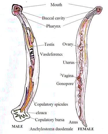
2. Body shape: It has a cylindrical, greyish-white body slightly bent at the anterior end. This develops a hook-like appearance.
3. Cuticle: The outer surface of a hookworm has a tough and flexible covering called cuticle. This cuticle protects hookworms from the host immune system.
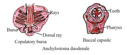
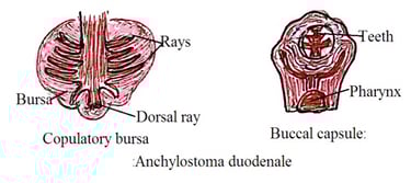
4. Oral apparatus: The anterior end of hookworm has a mouth. But it is not the terminal end. The mouth is surrounded by three pairs of teeth-like structures also called cutting plates. Four are hook-shaped and two are knob shaped. These cutting plates help to attach to the host intestine wall. It tears the intestinal mucous layer to get food.
5. Buccal capsule (Pharynx): It is the muscular part inside the mouth to suck blood from the host intestine wall.
6. Digestive system: It has a well-developed digestive system that starts from the buccal cavity and ends at the posterior end of the body. It does not have true anus. Thus, waste materials are excreted through the mouth. It has five digestive glands. Oesophageal gland secretion stops the clotting of blood inside the digestive system.
7. Reproductive system: Male and female hookworms exist individually. Male hookworm has curved posterior end. The curve end is called a copulatory bursa. The copulatory bursa attaches to the female hookworm during reproduction. Female hookworm has a straight posterior end. Female hookworm is larger in size than male hookworm.
Female hookworm produces and releases numerous eggs by internal fertilization. Fertilization occurs inside the intestine of female hookworms. These eggs are excreted from the host body with the stool.
Eggs of hookworms have the following features: They float on the saturated solution of sodium chloride.
1. Shape and size: Eggs are colorless, oval, or elliptical covered with a transparent membrane (hyaline). The membrane protects eggs against the harsh environment. Eggs do not cause infection in humans. 55 to 75 micrometers in length and 35 to 50 micrometers in width.
2. Contents: Eggs have embryos or developing larvae. Its larva can infect the host.

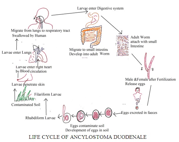
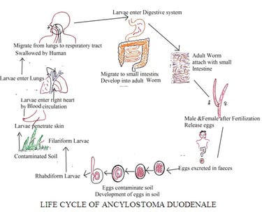
The Life Cycle of Ancylostoma (Hookworm)
Human is the definitive host (primary host) of hookworm. However, it requires the human body and a favorable environment outside the human body to complete its life cycle. It has only one host and does not require any intermediate host. The following are the stages of the hookworm life cycle.
Stage 1 (Eggs and the Environment): The infected human intestine has a female hookworm that releases numerous eggs. Eggs are excreted by the host with stool. These eggs contaminate the soil.
Stage 2 (Larvae development): Under favorable conditions (warm and moist soil), eggs release larvae. There are two stages of larva; Rhabditiform larva and Filariform larva.
Stage 3 (Infective stage): Filariform Larvae enter the human body through human skin. Normally, larva penetrates through bare feet and skin of other body parts in contact with contaminated soil. Larva can also enter the human body with contaminated food and water. But skin is the primary mode of infection. These larvae develop allergy and itching on the site entry into the skin. This is called (Ground itching or creeping eruption).
Stage 4 (Migration to the lungs): Filariform larvae enter human lungs with the blood. Larvae migrate from the lungs to the respiratory tract and are swallowed by the human during coughing. Swallowed larvae enter the human digestive system. Inside the human esophagus, it stays for about 10 days. During this period, the buccal capsule develops in hookworms. However, the buccal capsule does not have teeth (cutting plate). They enter the human small intestinal and develop into adult hookworms.
Stage 5 (Maturation and reproduction): It takes 3 to 4 weeks to develop into adult male and female hookworms inside intestine. They attach with human small intestine wall using their teeth. They feed on human blood and tissue fluid. Adult hookworms can survive inside human intestine for several years. Reproduction occurs and female hookworm produces eggs by internal fertilization. These eggs are excreted with the human stool.
Laboratory diagnosis of Ancylostoma (Hookworm):
Hookworm eggs and larvae are detected and identified in The laboratory to diagnose hookworm infection. The number of eggs present in the stool sample decides the intensity of hookworm infection. Administration of anthelmintic drugs promotes the excretion of worms in stool. Administration of anthelmintic drugs at bedtime and collection of stool samples in the morning is the most suitable technique to detect and identify parasites. This makes the detection easy in stool samples. The following methods are followed to diagnose hookworm infection in the medical laboratory.
1. Microscopic examination:
Direct wet method: A Fresh stool sample is mixed with the saline solution and the presence of hookworm eggs and larvae under the microscope is examined.
Several concentration techniques such as the sedimentation method, floating method, or formalin ether sedimentation techniques are used to concentrate the sample. Concentration techniques increase the chance of detection of hookworm eggs and larvae in the sample.
2. Stool culture: It is the most common and reliable method to detect and identify worms. A stool culture is used to cultivate hookworm larvae. A stool sample is inoculated into suitable culture media and incubated at a suitable temperature. After the incubation period, stool culture is examined under a microscope to identify hookworm larvae.
3. Serological test: Enzyme immunoassay (EIAs) is used in serological test to detect hookworm infection. It detects antibodies produced by the human body against hookworm antigens. It can detect chronic and acute hookworm infections.
4. Clinical diagnosis: Clinical symptoms such as anemia, GIT problem, and esophagus problem in conjunction with laboratory results confirm hookworm infection. Excretion of occult blood with stool along with anemia confirms the hookworm infection.
Dr Pramila Singh