
HSBTE Anatomy Physiology
HSBTE Question Paper DMLT Anatomy Physiology July 23. Solved Answer
HSBTE QUESTION SOLUTION
Dr Pramila Singh
11/24/202312 min read
HSBTE Question Paper Anatomy Physiology DMLT Examination July 23
1st Sem. Branch: DMLT
Subject: Anatomy & Physiology-I 221913
SECTION-A
Note: Multiple choice questions. All questions are compulsory (6x1=6)
.Q.1 The abdominal cavity contains the
a) Heart & Lung b) Liver & Spleen
c) Urinary bladder d) Urinary bladder& Lung Ans : b) Liver & Spleen
Q.2 The shoulder joint is
a) Nonmovable b) A synovial joint
c) Ball & socket joint d) None of above Ans: Ball & socket joint
Q.3 Cardiac muscles are ______ type of muscles
a) Voluntary b) Skeletal
c) Contractile d) Involuntary Ans: d) Involuntary.
Q.4 Full form of ECG is
a) Electro pathology b) Electro coronogram
c) Electrocardiogram d) None of above Ans: c) Electrocardiogram
Q.5 Lub Dub are the sounds of ___________
a) Liver b) Pulse
c) Lungs d) Heart Ans: d) Heart
Q.6 Which of the following is not part of the appendicular skeleton?
a) Scapula b) Ribs
c) Tibia d) Radius Ans: b) Ribs
SECTION-B
Note: Objective/ Completion type questions. All questions are compulsory. (6x1=6)
Q.7 What is meant by voluntary muscles?
Ans: The skeletal muscles that can be controlled by an individual will, are called skeletal muscles.
Q.8 Define BMR.
Ans: Basal Metabolic Rate (BMR) is the amount of energy an organism uses when the organism is at rest.
Q.9 Define Physiology.
Ans: Physiology is the study of functions of living organism organs.
Q.10 Expand AV & SV valves.
Ans: AV Valve: Atrio Ventricular. SV Valve: Valve Semilunar Valve.
Q.11 What are connective tissues?
Ans: Biological tissues that support, connect, and separate tissues are called connective tissues. Examples are bones, blood, cartilage, etc.
Q.12 Name the joints in the upper limb.
Ans: Sternoclavicular joints, Acromioclavicula, Shgoulder Joints, Elbow joints, Radioulnar joints,
SECTION-C
Note: Short answer type questions. Attempt any eight questions out of ten questions. (8x4=32)
Q.13 Write down the properties of muscular tissues.
Ans: PROPERTIES OF MUSCULAR TISSUES
All muscle cells have the following four common properties: Contractibility, excitability, extensibility, and elasticity.
1. Excitability: The ability of muscles to respond to a stimulus is the excitability property of muscle cells.
2. Contractibility: The ability of muscle cells to shorten is the property of muscle cells.
3. Elasticity: The ability of muscle cells to recoil to attain the original length after stretching is the elasticity property of muscle cells.
4. Extensibility: The ability of muscle cells to stretch is the extensibility property of muscle.
5. Conductivity: Stimulation in one part of muscle fibers spreads very quickly to other myofibrils.
6. Threshold: Contraction in muscle starts after receiving a certain intensity of stimulus. This is called threshold limit or threshold stimulus.
7. Muscle twitch: The contraction of a single muscle fiber in response to a single stimulus is called muscle twitch.
Q.14 Write a short note on the bones of the skull.
The Skull: It is a jointed, hardened framework of the head consisting of 29 bones. These 29 bones form four parts in the skull: Cranium, ear bone, Hyoid bone and facial bone.
1. Cranium: The cranium forms a the in the skull that protects the brain. There are eight cranial bones.
Frontal bone -1 Occipital bone-1 Sphenoid bone-1
Parietal bone-2 Temporal bone-2 Ethmoid bone-1
i. Frontal bone: The forehead and upper part of the orbital cavity is the frontal bone.
ii. Parietal bone: The side and roof of the skull are Parietal bone. The inner surface of the Parietal bone has deep furrows. Cranial arteries are present in these deep furrows.
iii. Occipital bone: It is present at the lower part and back of the cranial cavity. The occipital bone base has a large opening called the foramen magnum. The medulla oblongata of the brain passes through the foramen magnum and joins the spinal cord.
iv. Temporal bones: Temporal bones are present at each side of the skull.
v. Sphenoid bone: It looks like a bat with stretched-out wings.
vi. Ethmoid bone: It is cubical-shaped and light spongy bone situated at the nose roof.
2. Ear bones (ossicles): There are 6 ear bones. Three ear bones (ear ossicles) are found in each middle ear. One malleus. It is outer bone and hammer-shaped. One Incus, it is in middle bone and anvil-shaped. One stapes. It is the innermost stirrup-shaped bone. Stapes are the smallest bone of the body.
3. Hyoid bone: It is not bone of the skull.
4. Facial bone: There are 14 bones in the face. All are joined together by immovable joints called sutures except the mandibular bone. These bones are
· Nasal bone: Two in number forms Nose Bridge
· Palatine bones: Two in number, form mouth roof and nose floor.
· Lachrymal bones: Two in number, form tear duct.
· Zygomatic bones: Two in number, form cheekbone.
· Vomer bone: One in number, form the nose's lower part.
· Inferior turbinate bone: Two in number. form the upper jaw.
· Mandible: One in number forms the lower jaw. It is a skull-movable bone. It has a horse-shoe shape that carries lower teeth. It has outgrowth on each side
Maxilla: Two in number.
Q.15 Describe the mechanism of respiration.
Ans: MECHANISM OF BREATHING/Respiration: Breathing is the movement of fresh air into the lungs from the atmosphere and the movement of foul air out from the lungs into the atmosphere. There are two types of breathing: Inspiration and Expiration. Inspiration (inhalation) is the movement of fresh air into the lungs from the atmosphere. Expiration (exhalation) is the movement of foul air from the lungs into the atmosphere.
Phrenic muscles connect the diaphragm, ribs, and vertebral column. External intercostal muscles connect ribs. Internal intercostal muscles and abdominal muscles are used during forceful breathing.
Inspiration mechanism: Contraction of phrenic muscles changes the dome-shaped diaphragm into a straight diaphragm. This increases the thoracic cavity area. At the same time, intercostal muscles contract, and abdominal muscles relax. This causes the thorax to move outward and upward. This increases the thoracic cavity area. This helps to expand the lungs and generate negative pressure inside the lungs. To equalize the lung's negative pressure, air enters into lungs from the atmosphere.
Expiration mechanism: Relaxation of phrenic muscles brings the diaphragm to a normal position i.e. dome-shaped diaphragm. This decreases thoracic cavity volume. Relaxation of intercostal muscles brings ribs downward and inward. This also decreases thoracic cavity volume. At the same time, abdominal muscle contraction brings the abdominal organ upward. This again decreases thoracic cavity volume. This creates positive pressure inside the lungs. It forces air to rush out from the lungs into the atmosphere.
Route of exhaled air: Alveoli → bronchiole → bronchi → Trachea → Larynx → Pharynx → Nasal passage
Rate of breathing: In the adult, it is 12-24 per minute. In adolescents, it is 14-18 per minute. In children, it is 40-60 per minute.
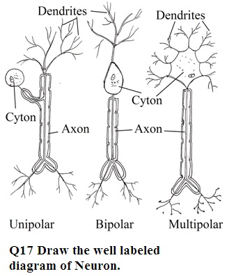
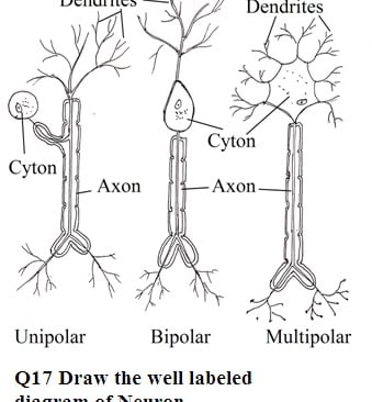
Q.16 Write down the working principle of ECG.
Ans: Basic Principle: ECG is a graphic record of electric current produced by cardiac muscle excitation. Cardiac muscle electrical excitation is due to cardiac impulse in the SA node, AV node, a bundle of HIS, and Purkinje fiber. An ECG instrument is used to record ECG. ECG shows 5 waves i.e. termed as PQRST.
Graphical Representation: The ECG wave consists of a P-wave, QRS-wave, and T-wave.
1. P wage represents electrical activity with the arterial contraction.
2. QRS Complex represents the electrical activity associated with ventricular contraction.
3. T wave represents the recovery (Repolarization) of the ventricles.
Q.18 Differentiate between the internal and external respiratory systems.
1. External respiration: It is also known as breathing. Breathing is a process to bring environmental oxygen into the body (inspiration) and bring out body carbon dioxide into the environment (expiration).
2. Internal respiration (Tissue Respiration): In internal respiration, cells consume oxygen in cellular metabolic activity to produce energy and carbon dioxide. Energy is utilized for biological activity.
Difference between respiration and breathing
Breathing is a Physical process. Respiration is a Biochemical process
BreathingUtilise energy. Respiration is a Release of energy
Breathing Exchange of gases between lungs and environment. Exchange of gases at the cellular level
Breathing is an Extracellular activity. Respiration is an Intracellular activity.
Breathing is an Extracellular activity, that does not involve enzyme Intra-cellular activity, Respiration is Catalysed by the enzyme.
Q.19 Describe the functional classification of joints.
Ans: The meeting point of two bones through the structural arrangement of tissues is called the joint of bones. These joints can be classified into the following types depending on their movability (Functional classification). These are
1. Synovial joints or Freely movable joints: Here one bone has a knob-like swelling while another bone has depression. Both knobs like swelling and depression structures are lined with smooth articular cartilages. Knob-like swelling of one bone fits into the Depression-like structure of another bone to form a synovial joint. Depression like the structure of bone is called a synovial cavity. Synovial fluid in synovial joints provides lubrication at joints to help in free movement in joints. Synovial joints are present in Shoulder joint, Elbow joint, wrist joint, hip joint, knee joint, and ankle joint.
2. Slightly movable or Cartilaginous Joints: (Intervertebral joint is an example of slightly movable joint). Thick elastic pad of fibro-cartilages is present at end of each bone. Bones join together through these pads of white fibro-cartilage. Examples are bone joints in the pelvic girdle (pubic symphysis), Joints between intervertebral discs (vertebrae).
3. Immovable joints or Fibrous Joint: There will be no movement between concerning bones. These bones join together by the presence of a strong bundle of white fibrous tissues (collagen fibers) at their end. Example: Joints among skull bones (These joints are called sutures), joints between teeth and maxilla, and joints between teeth and mandible.
Q.20 Define cardiac output. What determines cardiac output?
Ans: Cardiac output: The amount of blood pumped by the heart in one minute is called the cardiac output. A normal heart pumps 70 ml of blood on each ventricular contraction. The average heartbeat is 72 per minute. It means the heart pumps 72 X 70 ml of blood per minute which is 5.04 L. Cardiac output is almost equal to the total volume of blood present inside the body.
Q.21 Write the method of measuring blood pressure.
Measurement of Blood Pressure:
Apparatus: Sphygmomanometer: The sphygmomanometer consists of a cuff (inflatable bag), a compressible rubber bulb with an air pressure-releasing screw, and a mercury manometer. Two rubber tubes connect the rubber bulb, manometer, and inflatable bag (cuff). The manometer has mercury to measure pressure.
Procedure: The cuff is wrapped just above the elbow around the upper arm. A stethoscope diaphragm is kept on the brachial artery to hear a pulse. Air is pumped into the cuff by using a bulb connected with a stethoscope. Pumping air into a wrapped cuff will increase the level of mercury in the manometer of the sphygmomanometer.
Allow pressure to raise up to 200mm of Hg in the up manometer. At this pressure, blood will not pass through the brachial artery. There will be no radial pulse. Place the stethoscope diaphragm on the brachial artery of the bent elbow. Release air pressure by using a bulb screw vent. The release of air pressure allows blood to pass through the brachial artery. At a certain air pressure, the pulse will be felt. At this point, pressure in the mercury column of the manometer is noted. It is Systolic blood pressure. Pulse sound will disappear with further release of air. The pressure point of sound disappearance is called diastolic
If systolic blood pressure is more than 140 mm of Hg then it is called high blood pressure irrespective of diastolic blood pressure. Similarly, if diastolic blood pressure is more than 90 mm of Hg then it is considered as high blood pressure irrespective of systolic blood pressure. If systolic blood pressure is less than 90 mm of Hg then it is called low blood pressure irrespective of diastolic blood pressure. Similarly, if diastolic blood pressure is less than 60 mm of Hg it is considered as low blood pressure irrespective of systolic blood pressure.
Q.22 Differentiate between tendons and ligaments.
1. Ligaments are tough with elasticity. Tendons are tough with no elasticity
2. Ligaments joint and support between two bones. Tendon joint and support between bones and muscles.
3. Ligaments help bone movement. Tendons help bone and muscle movements.
4. Ligaments are compact fiber but not parallel. Tendons are compact and parallel fibres
SECTION-D
Note: Long answer type questions. Attempt any two questions out of three questions. (2x8=16)
Q.23 What are epithelial tissues? Give the classification of Epithelial tissues.
Epithelial tissues: Epithelial tissue consists of cells that grow on other cells or tissues. It covers both the external and internal surfaces of organs or body parts. Thus they form a protective covering on the external and internal surface of organ/body parts. They cover the body's surface eg. skin. They form lines on the upper and lower surface of hollow organs' Blood vessel walls.
Types of Epithelial tissues: There are two types of epithelial tissue
A. Simple epithelial tissue
B. Compound epithelial tissue
A. Simple epithelial tissue: It consists of a single layer of epithelial tissue on the basement membrane. Simple epithelial tissue is further subdivided depending on the shape of the cells.
a. Simple squamous epithelium tissue: It consists of large flat cells with flat nuclei. All cells are closely packed on the basement membrane.
b. Simple cuboidal epithelium: It consists of cuboidal, square-shaped cells with round nuclei. The nucleus is present in the centre of cells. Simple cuboidal epithelium present in ovaries and testes are called germinal epithelium cells. They produce ova in females and sperm in males.
c. Simple columnar epithelium: It consists of elongated cells having a column-like structure these column-shaped cells are present on the basement membrane. Cells have elongated nuclei away from the centre of cells. Simple columnar epithelium tissues are found in the internal lining of the stomach, intestine, rectum (internal lining of alimentary canal wall), Gall bladder, bile duct, and Respiratory tract wall.
d. Simple glandular epithelium: Columnar epithelium cells have glands that secrete juices and are called simple glandular epithelium. They are found in gastric glands, intestinal glands, and pancreatic lobules.
e. Simple ciliated epithelium: Cells of simple ciliated epithelial tissue may be columnar or cuboidal in shape. These cells have various fine hair-like structures called cilia on the free surface of cells.
f. Pseudo-stratified epithelium: It seems pseudostratified has a different layer but actually it has a single layer. Thus it is called pseudostratified epithelial tissues. It has two sizes of cells. There are two types of Pseudo-stratified epithelium.
Pseudostratified columnar epithelium
Pseudostratified columnar ciliated epithelium.
Both have column-shaped cells.
B. Compound Epithelial Tissue: Compound epithelial tissues consist of several layers of simple epithelial tissues. There are two types of compound epithelial tissues.
1. Stratified Epithelium
2. Transitional epithelium
Stratified Epithelium: It consists of several shapes of cells. Depending upon the shape of cells, stratified epithelium tissues are four types.
i. Stratified squamous epithelium
ii. Stratified cuboidal epithelium
iii. Stratified columnar epithelium
iv. Stratified ciliated columnar epithelium
Stratified squamous epithelium: It consists of several layers of cells. These layers can be divided into three parts superficial layer, Intermediate layer and Germinative layer.
The germinative layer is the innermost layer of this tissue. These cells may be column shape or cuboidal shape. The intermediate layer is the middle layer of this tissue. Outer layer is a superficial layer. It has flat cells and has elongated nuclei. Stratified squamous epithelium tissue is of two types.
Keratinized stratified squamous Epithelium.
Non Keratinized stratified squamous Epithelium.
Keratinized stratified squamous epithelium: Few outer layer cells have Keratin in place of cytoplasm. Keratin is a waterproof insoluble protein that gives hardness to cells. These are dead cells known as the horny layer or stratum corneum. It is found in skin epidermis.
Non-keratinized stratified squamous epithelium: They do not have keratin in cells in place of cytoplasm. Thus it is permeable to water and has moderate protection power. It is found in the buccal cavity, GIT, Urethra lower part, Vagina, Uterus lower part, cornea, etc.
Stratified cuboidal epithelium: There are two shapes of cells. Cells of the inner layer are columnar-shaped. Cells of the outer layer are cuboidal-shaped. It is found in the sweat gland, Pancreatic duct, and Salivary gland. etc
Stratified columnar epithelium: Outer layer cells and inner layer cells are columnar in shape. It is found in the urethra and mammary gland.
Stratified ciliated columnar epithelium: It consists of column-shaped cells both in outer layer and inner layer. Outer layer cells have cilia at the free surface. It i
Transitional Epithelium: cells of transitional epithelium tissues form 4 to 6 layers.
Innermost layer cells are columnar or cuboidal, Middle layer cells are polyhedral and superficial layer cells are globular. It does not have a basement membrane and germinative layer. But cell division by mitosis occurs. Cells have more elasticity than Stratified epithelium. Cells become flat after stretching. It is found in ureter, urinary bladder and inner part of the urethra. They can be stretched without any damage. It acts as protective.
Q.24 Explain about the following
a) Tidal volume
Tidal volume: The volume of air that enters into the lungs and comes out from the lungs in normal breathing without any extra force is called tidal volume or tidal air. The volume of tidal air is 500 ml. 350 ml of tidal air remains in the respiratory zone and 150 ml of tidal air remains in conducting. This 150 ml does not participate in pulmonary respiration (Exchange of gases in lungs). 150 ml of tidal air is called dead space air. Lungs' total air capacity is 4.5 litres to 5 litres. The tidal air is one by a tenth of the total air capacity of the lungs.
b) Vital capacity
Vital capacity: The total volume of air that enters into the lungs and comes out from the lungs by forceful breathing is called vital capacity. Vital capacity depends upon age, sex, height, physical activity etc. In a normal man, a vital capacity of air is 4 to 5 litres and in normal women, it is 3 to 4 litres. It is measured in Spirometry.
Dr Pramila Singh