Nucleotide Metabolism and Purine Pyrimidine Biosynthesis
Practice MCQs on Nucleotide Metabolism and Purine Pyrimidine Biosynthesis gout, hyperuricemia, genome structure, DNA replication, transcription, and translation for GPAT, NIPER, and pharmacist exams.
Dr. Alok Singh
7/23/202614 min read
MCQs: Nucleotide Metabolism, Gout, Genome Structure & Central Dogma
Quick Revision Notes for GPAT, NIPER, AIIMS Pharmacist, Railway Pharmacist, SSC, ESIC & State Pharmacist Exams
Nucleotides are essential biomolecules that serve as the building blocks of DNA and RNA. They also participate in energy transfer, cellular signaling, and several metabolic reactions. The metabolism of nucleotides includes their biosynthesis, salvage, and degradation.
For competitive examinations, special attention should be given to purine and pyrimidine synthesis, uric acid formation, gout, Lesch-Nyhan syndrome, orotic aciduria, SCID, DNA replication, transcription, translation, and chromatin organization.
1. Nucleotide Structure and Functions
A nucleotide consists of three components:
Nitrogenous base + Pentose sugar + Phosphate
Nitrogenous Bases
Purines:
Adenine (A)
Guanine (G)
Pyrimidines:
Cytosine (C)
Thymine (T)
Uracil (U)
Easy Memory Trick
"Pure As Gold" → Purines = Adenine + Guanine
"CUT the PY" → Pyrimidines = Cytosine + Uracil + Thymine
Nucleoside vs Nucleotide
Nucleoside = Base + Sugar
Nucleotide = Base + Sugar + Phosphate
Important Functions
Nucleotides are involved in:
Formation of DNA and RNA
Energy transfer: ATP and GTP
Cell signaling: cAMP and cGMP
Coenzyme structure: NAD⁺, FAD, and CoA contain nucleotide components
Activation of metabolic intermediates
2. Purine Nucleotide Biosynthesis
Purine nucleotides are synthesized by two major pathways:
De novo synthesis
Salvage pathway
A. De Novo Purine Synthesis
In de novo synthesis, the purine ring is gradually constructed directly on a ribose phosphate molecule.
The starting material is:
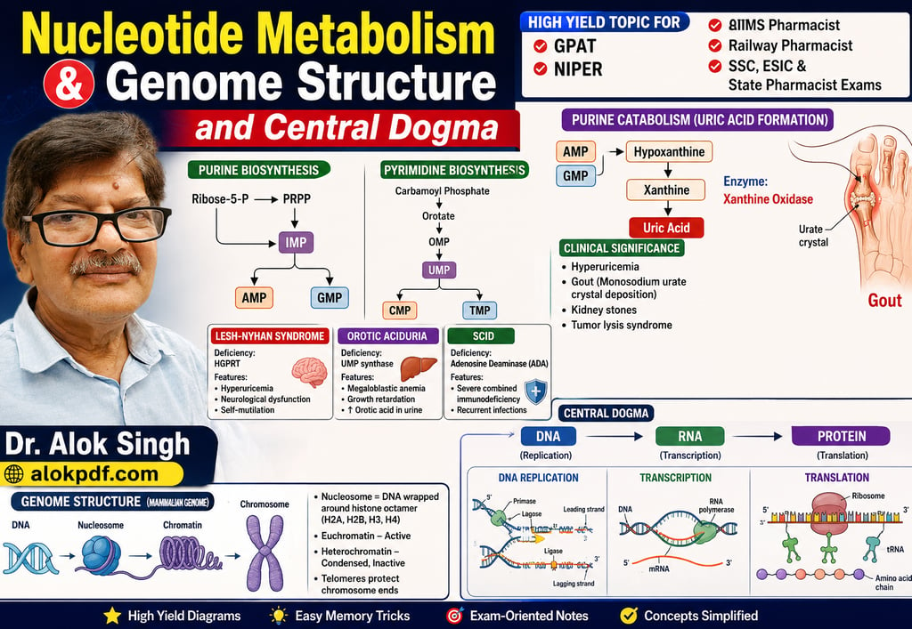
Ribose-5-phosphate → PRPP → IMP → AMP/GMP
The first fully formed purine nucleotide is IMP (inosine monophosphate).
IMP serves as the common precursor for:
AMP (adenosine monophosphate)
GMP (guanosine monophosphate)
Easy Memory Trick
"IMP is the IMPortant junction"
IMP → AMP or GMP
Important Point
The purine ring is built on PRPP.
This is a very common competitive-exam question.
Rate-Limiting/Committed Step
PRPP → 5-Phosphoribosylamine
Enzyme:
Glutamine-PRPP amidotransferase
It is inhibited by the end products:
IMP, AMP, and GMP
Thus, purine synthesis is regulated by feedback inhibition.
3. Purine Salvage Pathway
The salvage pathway recycles free purine bases to form nucleotides.
Important enzymes:
HGPRT
Hypoxanthine + PRPP → IMP
Guanine + PRPP → GMP
APRT
Adenine + PRPP → AMP
Clinical Importance: Lesch-Nyhan Syndrome
Complete deficiency of HGPRT causes Lesch-Nyhan syndrome.
Important features include:
Severe hyperuricemia
Gout-like symptoms
Neurological dysfunction
Dystonia
Intellectual disability
Characteristic self-injurious behavior
Easy Memory Trick
"HGPRT → HyperGout + Psychiatric/neurological Problems + Repeated self-injury"
Remember:
HGPRT deficiency = Lesch-Nyhan syndrome
4. Pyrimidine Nucleotide Biosynthesis
The major pyrimidine nucleotides are:
CMP
UMP
TMP
Unlike purines, the pyrimidine ring is synthesized first and then attached to PRPP.
Easy Comparison
Purines: Ring built on PRPP
Pyrimidines: Ring built first, then attached to PRPP
The pathway can be summarized as:
Carbamoyl phosphate → Orotate → OMP → UMP
UMP is the central precursor for other pyrimidine nucleotides.
Important Enzyme
Carbamoyl phosphate synthetase II (CPS-II)
Location:
Cytoplasm
Function:
Produces carbamoyl phosphate for pyrimidine synthesis.
High-Yield Difference
CPS-I
Mitochondria
Urea cycle
Uses ammonia
CPS-II
Cytoplasm
Pyrimidine synthesis
Uses glutamine as nitrogen donor
Easy Memory Trick
"CPS-I = I am for urea"
"CPS-II = I make pyrimidines"
5. Orotic Aciduria
Orotic aciduria is caused by deficiency of:
UMP synthase
This enzyme is required for conversion of orotic acid to UMP.
Features
Increased urinary orotic acid
Megaloblastic anemia
Growth retardation
No significant hyperammonemia
Treatment
Uridine supplementation
Uridine bypasses the metabolic block and restores pyrimidine nucleotide synthesis.
Exam Trap
Orotic aciduria → UMP synthase deficiency
Do not confuse it with OTC deficiency, which also causes increased orotic acid but is associated with hyperammonemia.
6. Catabolism of Purine Nucleotides
Purine degradation ultimately produces uric acid in humans.
Adenine Pathway
AMP → Adenosine → Inosine → Hypoxanthine → Xanthine → Uric acid
Guanine Pathway
GMP → Guanosine → Guanine → Xanthine → Uric acid
The enzyme xanthine oxidase catalyzes:
Hypoxanthine → Xanthine
and
Xanthine → Uric acid
Easy Memory Trick
"HX → X → UA"
Hypoxanthine → Xanthine → Uric Acid
Why Do Humans Produce Uric Acid?
Humans lack functional uricase, the enzyme that converts uric acid into the more soluble compound allantoin.
Therefore:
Purines → Uric acid = Final major product in humans
7. Hyperuricemia
Hyperuricemia means an abnormally increased concentration of uric acid in the blood.
It may occur due to:
1. Increased Uric Acid Production
Examples:
Increased purine breakdown
High purine intake
Rapid cell destruction
Tumor lysis syndrome
Certain genetic disorders
2. Decreased Uric Acid Excretion
This is a common cause of hyperuricemia.
It may occur due to:
Renal impairment
Reduced renal urate excretion
Certain medications
Clinical Consequences
Persistent hyperuricemia may lead to:
Gout
Urate crystal deposition
Kidney stones
Uric acid nephropathy
8. Gout
Gout is an inflammatory disorder caused by deposition of monosodium urate crystals in joints and tissues.
Classic Features
Severe joint pain
Swelling
Redness
Inflammation
Often begins in the first metatarsophalangeal joint
This is called:
Podagra
Pathogenesis
Hyperuricemia → Monosodium urate crystal deposition → Inflammation → Gout
Urate crystals activate inflammatory pathways and attract neutrophils, producing intense inflammation.
9. Important Drugs Used in Gout
A. Allopurinol
Mechanism:
Xanthine oxidase inhibitor
It decreases:
Hypoxanthine → Xanthine → Uric acid
Used mainly for long-term control of hyperuricemia.
Remember:
Allopurinol = Allopurine inhibits Xanthine Oxidase
B. Febuxostat
Selective xanthine oxidase inhibitor
Reduces uric acid synthesis
Remember:
Allopurinol + Febuxostat → Decrease uric acid production
C. Probenecid
Uricosuric drug
Increases renal excretion of uric acid
Remember:
Probenecid → Promotes urate excretion
D. Colchicine
Colchicine does not primarily lower uric acid levels.
It reduces inflammation by inhibiting microtubule polymerization and decreasing neutrophil migration.
It is particularly useful in acute gout attacks.
Important Exam Comparison
Drug Main Action
Allopurinol Inhibits xanthine oxidase
Febuxostat Inhibits xanthine oxidase
Probenecid Increases uric acid excretion
Colchicine Reduces inflammation in acute gout
10. Tumor Lysis Syndrome and Hyperuricemia
Rapid destruction of cancer cells can release large quantities of nucleic acids.
The sequence is:
Rapid cell destruction → Purine release → Purine degradation → Increased uric acid → Hyperuricemia
This may result in:
Acute kidney injury
Uric acid nephropathy
Electrolyte abnormalities
Therefore, tumor lysis syndrome is an important clinical cause of acute hyperuricemia.
11. Genome Structure and Organization
The genome is the complete genetic material of an organism.
In mammals, the genome is mainly located in the nucleus, with a small amount of DNA present in mitochondria.
Mammalian Nuclear Genome
DNA is organized into:
DNA → Nucleosomes → Chromatin → Chromosomes
Nucleosome
The nucleosome is the basic structural unit of chromatin.
It consists of DNA wrapped around a core of eight histone proteins:
2 × H2A
2 × H2B
2 × H3
2 × H4
Histone H1 binds linker DNA between nucleosomes.
Easy Memory Trick
"The 4 Histones Make an 8"
H2A + H2B + H3 + H4, each present twice = 8 histone proteins
12. Euchromatin and Heterochromatin
Euchromatin
Less condensed
Transcriptionally active
Accessible to transcription machinery
Heterochromatin
Highly condensed
Generally transcriptionally inactive or less active
Easy Memory Trick
EU = Expressed
HETERO = Heavily packed
13. Chromosomes and Telomeres
Chromosomes contain DNA and associated proteins.
Important chromosome regions include:
Centromere: Important for chromosome segregation
Telomeres: Protect chromosome ends
Telomeres contain repetitive DNA sequences and prevent:
Chromosome degradation
End-to-end chromosome fusion
The enzyme telomerase maintains telomere length.
High-Yield Point
Telomerase activity is increased in:
Germ cells
Stem cells
Many cancer cells
14. Central Dogma of Molecular Biology
The central dogma describes the flow of genetic information:
DNA → RNA → Protein
The three major processes are:
DNA Replication: DNA → DNA
Transcription: DNA → RNA
Translation: RNA → Protein
Easy Memory Trick
"Copy → Write → Translate"
Replication = Copy DNA
Transcription = Write RNA
Translation = Make Protein
15. DNA Replication
DNA replication is the process by which DNA makes an identical copy of itself.
It occurs during the S phase of the cell cycle.
Important Characteristics
DNA replication is:
Semiconservative
Bidirectional
Template-directed
Semiconservative Replication
Each daughter DNA molecule contains:
One parental strand + One newly synthesized strand
Important Enzymes
Helicase
→ Unwinds DNA double helix
Primase
→ Synthesizes RNA primer
DNA polymerase
→ Synthesizes new DNA
DNA ligase
→ Joins DNA fragments
Topoisomerase
→ Relieves DNA supercoiling
Direction of DNA Synthesis
DNA polymerase synthesizes DNA only in the:
5′ → 3′ direction
16. Leading and Lagging Strands
Leading Strand
Synthesized continuously
Requires one RNA primer
Lagging Strand
Synthesized discontinuously
Forms Okazaki fragments
Requires multiple RNA primers
DNA ligase joins the Okazaki fragments.
Easy Memory Trick
"Leading = Continuous"
"Lagging = Little pieces"
17. Transcription
Transcription is the synthesis of RNA using DNA as a template.
DNA → RNA
In eukaryotic cells, transcription mainly occurs in the nucleus.
Important RNA Polymerases
RNA Polymerase I
→ Major rRNA synthesis
RNA Polymerase II
→ mRNA synthesis
RNA Polymerase III
→ tRNA and 5S rRNA synthesis
Easy Memory Trick
"I = rRNA, II = mRNA, III = tRNA"
18. Eukaryotic mRNA Processing
The primary RNA transcript undergoes processing before becoming mature mRNA.
Important steps:
1. 5′ Capping
A 7-methylguanosine cap is added to the 5′ end.
Functions:
Protects mRNA from degradation
Helps nuclear export
Helps translation initiation
2. Splicing
Introns are removed and exons are joined.
Splicing is carried out by the spliceosome.
3. Polyadenylation
A poly-A tail is added to the 3′ end.
Functions:
Increases mRNA stability
Protects mRNA from degradation
Helps efficient translation
Easy Memory Trick
"Cap → Cut → Tail"
5′ Cap → Remove introns → 3′ Poly-A tail
19. Translation
Translation is the process by which the information in mRNA is used to synthesize a protein.
mRNA → Protein
It occurs on ribosomes.
Three Main Stages
Initiation
Elongation
Termination
Initiation Codon
AUG
AUG generally codes for:
Methionine
In bacteria, the initiating amino acid is:
N-formylmethionine (fMet)
Stop Codons
UAA
UAG
UGA
These do not code for amino acids.
Easy Memory Trick
"AUG = Start"
"UAA, UAG, UGA = Stop"
20. tRNA and Anticodon
Transfer RNA (tRNA) carries amino acids to the ribosome.
Each tRNA contains an anticodon that pairs with the complementary codon on mRNA.
Remember:
mRNA = Codon
tRNA = Anticodon
The ribosome reads the mRNA codons and links amino acids together through peptide bonds.
The catalytic activity responsible for peptide bond formation is primarily associated with ribosomal RNA (rRNA).
Thus, the ribosome functions as a ribozyme.
21. High-Yield Comparison: Replication vs Transcription vs Translation
Feature Replication Transcription Translation
Basic process DNA → DNA DNA → RNA RNA → Protein
Main product DNA RNA Protein
Main machinery DNA polymerase RNA polymerase Ribosome
Template DNA DNA mRNA
Main location in eukaryotes Nucleus Nucleus Cytoplasm/Rough ER
Primer required? Yes No No
Direction 5′→3′ 5′→3′ mRNA read 5′→3′
22. Must-Remember Clinical and Exam Associations
Purine Metabolism
HGPRT deficiency
→ Lesch-Nyhan syndrome
ADA deficiency
→ Severe combined immunodeficiency (SCID)
Xanthine oxidase
→ Hypoxanthine → Xanthine → Uric acid
Uricase absent in humans
→ Uric acid is the final major product of purine degradation
Allopurinol / Febuxostat
→ Xanthine oxidase inhibitors
Probenecid
→ Uricosuric drug
Colchicine
→ Reduces inflammation in acute gout
Pyrimidine Metabolism
UMP synthase deficiency
→ Orotic aciduria
Orotic aciduria
→ Megaloblastic anemia + increased urinary orotic acid
Treatment
→ Uridine
CPS-II
→ Pyrimidine synthesis
CPS-I
→ Urea cycle
Genome and Central Dogma
Nucleosome
→ Basic unit of chromatin
H1
→ Linker DNA
Euchromatin
→ Active transcription
Heterochromatin
→ Condensed and relatively inactive
Telomere
→ Protects chromosome ends
Helicase
→ Unwinds DNA
Primase
→ Makes RNA primer
DNA ligase
→ Joins Okazaki fragments
RNA polymerase II
→ mRNA synthesis
Spliceosome
→ Removes introns
AUG
→ Start codon
UAA, UAG, UGA
→ Stop codons
Final Rapid Revision Formula
Purine
PRPP → IMP → AMP/GMP → Uric Acid
Pyrimidine
Carbamoyl phosphate → Orotate → UMP → Other pyrimidines
Gout
↑ Uric Acid → Urate Crystals → Inflammation
Lesch-Nyhan
↓ HGPRT → ↑ Purine degradation → ↑ Uric Acid
Orotic Aciduria
↓ UMP Synthase → ↑ Orotic Acid → Megaloblastic Anemia
Central Dogma
DNA → RNA → Protein
DNA Replication
Helicase → Primase → DNA Polymerase → Ligase
mRNA Processing
5′ Cap → Splicing → 3′ Poly-A Tail
Translation
AUG → Initiation → Elongation → Stop Codon
One-Line Exam Takeaways
Purines are built on PRPP; pyrimidine rings are built first.
IMP is the common precursor of AMP and GMP.
HGPRT deficiency causes Lesch-Nyhan syndrome.
ADA deficiency causes SCID.
UMP synthase deficiency causes hereditary orotic aciduria.
Xanthine oxidase converts hypoxanthine to xanthine and xanthine to uric acid.
Allopurinol and febuxostat inhibit xanthine oxidase.
Probenecid increases uric acid excretion.
Colchicine reduces inflammation in acute gout.
A nucleosome is the basic unit of chromatin.
Euchromatin is transcriptionally active.
DNA replication is semiconservative, and DNA synthesis occurs 5′→3′.
RNA polymerase II synthesizes mRNA.
Spliceosomes remove introns.
AUG is the start codon; UAA, UAG, and UGA are stop codons.
The central dogma is DNA → RNA → Protein.
Below is an MCQ set designed in the style of questions commonly encountered in GPAT, NIPER, AIIMS Pharmacist, Railway Pharmacist, SSC, ESIC, and State Pharmacist examinations.
1. A patient with severe gout is prescribed allopurinol. The primary biochemical basis for its therapeutic action is inhibition of:
A. Adenylosuccinate synthetase
B. Xanthine oxidase
C. Hypoxanthine-guanine phosphoribosyltransferase
D. Carbamoyl phosphate synthetase II
Answer: B. Xanthine oxidase
Explanation: Allopurinol is a structural analogue of hypoxanthine and inhibits xanthine oxidase, reducing the conversion of hypoxanthine → xanthine → uric acid. It is therefore useful in chronic hyperuricemia and gout.
2. A child presents with self-mutilation, dystonia, and hyperuricemia. The most likely enzyme deficiency is:
A. Adenosine deaminase
B. Hypoxanthine-guanine phosphoribosyltransferase
C. Xanthine oxidase
D. Thymidylate synthase
Answer: B. Hypoxanthine-guanine phosphoribosyltransferase (HGPRT)
Explanation: Complete HGPRT deficiency causes Lesch-Nyhan syndrome. Defective purine salvage increases PRPP and decreases IMP/GMP formation, stimulating de novo purine synthesis and increasing uric acid production.
3. Which reaction represents a key regulatory step in de novo purine nucleotide synthesis?
A. IMP → AMP
B. Ribose-5-phosphate → PRPP
C. PRPP → 5-Phosphoribosylamine
D. GMP → Uric acid
Answer: C. PRPP → 5-Phosphoribosylamine
Explanation: Glutamine-PRPP amidotransferase catalyzes the committed step of de novo purine synthesis. It is inhibited by purine nucleotides such as IMP, AMP, and GMP.
4. Which statement correctly distinguishes purine and pyrimidine biosynthesis?
A. The purine ring is synthesized first and then attached to ribose
B. The pyrimidine ring is synthesized directly on PRPP
C. The purine ring is assembled stepwise on a ribose phosphate scaffold
D. Both purine and pyrimidine rings are synthesized exclusively in mitochondria
Answer: C. The purine ring is assembled stepwise on a ribose phosphate scaffold
Explanation: In purine synthesis, the ring is built sequentially on PRPP. In pyrimidine synthesis, the pyrimidine ring is synthesized first and subsequently attached to PRPP.
5. The immediate precursor of uric acid in purine catabolism is:
A. Adenine
B. Hypoxanthine
C. Xanthine
D. Guanine
Answer: C. Xanthine
Explanation: Purine degradation proceeds through hypoxanthine → xanthine → uric acid, with xanthine oxidase catalyzing both oxidation steps.
6. A patient receiving 6-mercaptopurine develops severe myelosuppression after starting azathioprine. Which enzyme is most important for the metabolism of these drugs?
A. Xanthine oxidase
B. HGPRT
C. Dihydrofolate reductase
D. Thymidine kinase
Answer: B. HGPRT
Explanation: 6-Mercaptopurine is activated by HGPRT to nucleotide derivatives that inhibit purine synthesis. Its metabolism also involves xanthine oxidase and TPMT, making drug interactions clinically important.
7. Which enzyme catalyzes the conversion of ribonucleotides into deoxyribonucleotides?
A. DNA polymerase
B. Ribonucleotide reductase
C. RNA polymerase
D. Thymidylate synthase
Answer: B. Ribonucleotide reductase
Explanation: Ribonucleotide reductase converts ribonucleoside diphosphates into deoxyribonucleotides, providing precursors required for DNA synthesis.
8. A patient with gout is started on febuxostat. Compared with allopurinol, febuxostat primarily acts by:
A. Inhibiting HGPRT
B. Inhibiting xanthine oxidase
C. Increasing uric acid excretion by inhibiting URAT1
D. Inhibiting purine salvage
Answer: B. Inhibiting xanthine oxidase
Explanation: Febuxostat is a selective xanthine oxidase inhibitor. It reduces uric acid formation and is used for chronic hyperuricemia associated with gout.
9. The major end product of purine catabolism in humans is uric acid because humans lack:
A. Xanthine oxidase
B. Adenosine deaminase
C. Uricase
D. HGPRT
Answer: C. Uricase
Explanation: Humans lack functional uricase, the enzyme that converts uric acid to the more soluble allantoin. Consequently, uric acid accumulates as the final product of purine degradation.
10. Which condition is most directly associated with deposition of monosodium urate crystals in joints?
A. Rheumatoid arthritis
B. Osteoarthritis
C. Gout
D. Osteoporosis
Answer: C. Gout
Explanation: Gout results from deposition of needle-shaped monosodium urate crystals in joints and tissues due to hyperuricemia, triggering intense inflammatory responses.
11. A patient develops acute gout after chemotherapy for leukemia. The most likely cause is:
A. Increased pyrimidine synthesis
B. Tumor lysis syndrome
C. Increased urea cycle activity
D. Reduced DNA degradation
Answer: B. Tumor lysis syndrome
Explanation: Rapid destruction of tumor cells releases nucleic acids, increasing purine degradation and uric acid production. This can cause acute hyperuricemia and urate nephropathy.
12. Which drug promotes uric acid excretion primarily by inhibiting renal tubular urate reabsorption?
A. Probenecid
B. Allopurinol
C. Colchicine
D. Febuxostat
Answer: A. Probenecid
Explanation: Probenecid is a uricosuric agent that reduces renal tubular reabsorption of uric acid, increasing urinary urate excretion.
13. A deficiency of adenosine deaminase is classically associated with:
A. Lesch-Nyhan syndrome
B. Severe combined immunodeficiency
C. Gout
D. Orotic aciduria
Answer: B. Severe combined immunodeficiency (SCID)
Explanation: ADA deficiency causes accumulation of deoxyadenosine and dATP, which inhibits ribonucleotide reductase and impairs lymphocyte development, producing SCID.
14. Orotic aciduria is most directly associated with deficiency of:
A. UMP synthase
B. HGPRT
C. Adenosine deaminase
D. Xanthine oxidase
Answer: A. UMP synthase
Explanation: UMP synthase deficiency causes hereditary orotic aciduria. Orotic acid accumulates, resulting in megaloblastic anemia and growth retardation without hyperammonemia.
15. Which intermediate is common to the biosynthesis of both AMP and GMP?
A. UMP
B. IMP
C. PRPP
D. Carbamoyl phosphate
Answer: B. IMP
Explanation: IMP (inosine monophosphate) is the first fully formed purine nucleotide and serves as the precursor for both AMP and GMP.
16. In mammalian cells, the majority of genomic DNA is located in:
A. Ribosomes
B. Mitochondria
C. Nucleus
D. Lysosomes
Answer: C. Nucleus
Explanation: Most mammalian genomic DNA is located in the nucleus, organized into chromosomes. A small, distinct genome is also present in mitochondria.
17. The fundamental repeating unit of chromatin is:
A. Centrosome
B. Nucleosome
C. Spliceosome
D. Ribosome
Answer: B. Nucleosome
Explanation: A nucleosome consists of approximately 147 base pairs of DNA wrapped around a histone octamer containing H2A, H2B, H3, and H4.
18. Which histone protein is primarily associated with linker DNA between nucleosomes?
A. H1
B. H2A
C. H3
D. H4
Answer: A. H1
Explanation: Histone H1 binds linker DNA and promotes higher-order chromatin organization.
19. Which form of chromatin is generally transcriptionally active?
A. Heterochromatin
B. Euchromatin
C. Centromeric chromatin
D. Constitutive heterochromatin
Answer: B. Euchromatin
Explanation: Euchromatin is relatively less condensed and generally accessible to transcription machinery. Heterochromatin is more condensed and transcriptionally inactive or less active.
20. Telomeres are important because they:
A. Initiate transcription
B. Protect chromosome ends from degradation and fusion
C. Encode ribosomal proteins
D. Initiate translation
Answer: B. Protect chromosome ends from degradation and fusion
Explanation: Telomeres consist of repetitive DNA sequences that protect the ends of chromosomes. Telomerase maintains telomere length in germ cells, stem cells, and many cancer cells.
21. DNA replication is described as semiconservative because:
A. Only one strand is copied
B. Each daughter DNA molecule contains one parental and one newly synthesized strand
C. Both daughter strands are newly synthesized
D. DNA replication occurs only during mitosis
Answer: B. Each daughter DNA molecule contains one parental and one newly synthesized strand
Explanation: In semiconservative replication, each daughter DNA molecule contains one original parental strand and one newly synthesized complementary strand.
22. Which enzyme synthesizes the RNA primer required for DNA replication?
A. DNA polymerase
B. DNA ligase
C. Primase
D. Topoisomerase
Answer: C. Primase
Explanation: Primase, an RNA polymerase, synthesizes short RNA primers that provide the free 3′-OH group required by DNA polymerase.
23. The major difference between leading and lagging strand synthesis is that:
A. Leading strand synthesis is discontinuous
B. Lagging strand synthesis is discontinuous
C. Both are synthesized independently of primers
D. Lagging strand synthesis occurs in the 3′→5′ direction
Answer: B. Lagging strand synthesis is discontinuous
Explanation: DNA polymerase synthesizes DNA only in the 5′→3′ direction. Therefore, the lagging strand is synthesized discontinuously as Okazaki fragments.
24. Which enzyme joins Okazaki fragments?
A. DNA helicase
B. DNA ligase
C. Primase
D. Topoisomerase II
Answer: B. DNA ligase
Explanation: DNA ligase seals nicks in the sugar-phosphate backbone by forming phosphodiester bonds between adjacent DNA fragments.
25. A drug inhibits DNA topoisomerase II. Which process is most directly affected?
A. Removal of supercoiling during DNA replication
B. Formation of peptide bonds
C. RNA splicing
D. Amino acid activation
Answer: A. Removal of supercoiling during DNA replication
Explanation: Topoisomerases control DNA supercoiling. Topoisomerase II introduces transient double-strand breaks to relieve torsional stress and separate intertwined DNA molecules.
26. The enzyme responsible for synthesis of messenger RNA in eukaryotic cells is:
A. RNA polymerase I
B. RNA polymerase II
C. RNA polymerase III
D. DNA polymerase α
Answer: B. RNA polymerase II
Explanation: RNA polymerase II synthesizes precursor mRNA and several small nuclear RNAs. RNA polymerase I mainly synthesizes rRNA, while RNA polymerase III synthesizes tRNA and 5S rRNA.
27. Which modification is added to the 5′ end of eukaryotic mRNA?
A. Poly-A tail
B. 7-Methylguanosine cap
C. Intron
D. Anticodon
Answer: B. 7-Methylguanosine cap
Explanation: The 5′ cap protects mRNA from degradation and assists in nuclear export and translation initiation.
28. The poly-A tail of eukaryotic mRNA is added to the:
A. 5′ end
B. 3′ end
C. Middle of the transcript
D. Promoter region
Answer: B. 3′ end
Explanation: The poly-A tail is added to the 3′ end of most eukaryotic mRNAs. It contributes to mRNA stability and efficient translation.
29. Introns are removed from eukaryotic pre-mRNA by:
A. Ribosomes
B. Spliceosomes
C. DNA polymerases
D. Proteasomes
Answer: B. Spliceosomes
Explanation: Spliceosomes, composed of small nuclear RNAs and proteins, remove introns and join exons during RNA splicing.
30. Which sequence is most directly involved in initiating transcription?
A. Promoter
B. Anticodon
C. Poly-A tail
D. Stop codon
Answer: A. Promoter
Explanation: The promoter is a DNA sequence recognized by transcription machinery and transcription factors, determining where RNA synthesis begins.
31. During translation, the anticodon of tRNA pairs with the:
A. Promoter
B. Coding strand of DNA
C. Codon of mRNA
D. Ribosomal RNA
Answer: C. Codon of mRNA
Explanation: The tRNA anticodon base-pairs with the complementary mRNA codon, ensuring incorporation of the correct amino acid into the growing polypeptide.
32. The universal initiation codon for translation is:
A. UAA
B. AUG
C. UGA
D. UAG
Answer: B. AUG
Explanation: AUG generally serves as the start codon and codes for methionine. In bacteria, the initiating amino acid is formylmethionine (fMet).
33. Which of the following is a termination codon?
A. AUG
B. UGG
C. UGA
D. GUG
Answer: C. UGA
Explanation: UGA, along with UAA and UAG, is a stop codon. These codons do not specify amino acids and signal termination of translation.
34. The catalytic activity responsible for peptide bond formation is primarily associated with:
A. DNA
B. Ribosomal RNA
C. Transfer RNA
D. Messenger RNA
Answer: B. Ribosomal RNA
Explanation: The ribosome functions as a ribozyme. Its peptidyl transferase activity is primarily associated with rRNA in the large ribosomal subunit.
35. A mutation changes a codon for an amino acid into UAA. This type of mutation is called:
A. Missense mutation
B. Silent mutation
C. Nonsense mutation
D. Frameshift mutation
Answer: C. Nonsense mutation
Explanation: A nonsense mutation converts an amino acid codon into a premature stop codon, resulting in early termination of protein synthesis.
36. Which sequence correctly represents the central dogma of molecular biology?
A. RNA → DNA → Protein
B. DNA → RNA → Protein
C. Protein → RNA → DNA
D. DNA → Protein → RNA
Answer: B. DNA → RNA → Protein
Explanation: The classical central dogma describes information flow from DNA to RNA by transcription and from RNA to protein by translation.
37. A mutation occurs in the promoter region of a gene but leaves the coding sequence intact. The most likely consequence is:
A. Altered amino acid sequence
B. Altered transcriptional regulation
C. Altered genetic code
D. Direct alteration of ribosomal structure
Answer: B. Altered transcriptional regulation
Explanation: A promoter controls transcription initiation. A mutation may reduce or increase gene expression without necessarily changing the amino acid sequence of the encoded protein.
38. Which of the following correctly pairs the process with its cellular location in a typical eukaryotic cell?
A. Transcription—cytoplasm; translation—nucleus
B. Transcription—nucleus; translation—cytoplasm
C. Both transcription and translation—nucleus
D. Both transcription and translation—mitochondria
Answer: B. Transcription—nucleus; translation—cytoplasm
Explanation: In eukaryotic cells, transcription occurs mainly in the nucleus, whereas translation occurs on cytoplasmic ribosomes or ribosomes associated with rough ER.
39. A researcher observes that a gene is highly expressed despite its DNA being relatively accessible and less condensed. This DNA is most likely present as:
A. Heterochromatin
B. Euchromatin
C. Centromeric DNA
D. Telomeric DNA
Answer: B. Euchromatin
Explanation: Euchromatin is less condensed and generally more accessible to transcription factors and RNA polymerase, favoring active gene expression.
40. Which combination is correctly matched?
A. Allopurinol—Inhibits HGPRT
B. Probenecid—Xanthine oxidase inhibitor
C. Colchicine—Reduces neutrophil-mediated inflammation in acute gout
D. ADA deficiency—Hyperuricemia as the primary clinical feature
Answer: C. Colchicine—Reduces neutrophil-mediated inflammation in acute gout
Explanation: Colchicine inhibits microtubule polymerization and reduces neutrophil migration and inflammatory responses to urate crystals. Allopurinol inhibits xanthine oxidase, while ADA deficiency is classically associated with SCID.
Quick Revision: High-Yield Exam Traps
Topic Key Point
Purine synthesis Purine ring is built on PRPP
Pyrimidine synthesis Ring is synthesized before attachment to PRPP
IMP Common precursor of AMP and GMPH
GPRT deficiency Lesch-Nyhan syndrome
ADA deficiency SCID
UMP synthase deficiency Orotic aciduria
Xanthine oxidase Hypoxanthine → Xanthine → Uric acid
Allopurinol/Febuxostat Xanthine oxidase inhibitors
Probenecid Uricosuric drug
Colchicine Controls inflammation in acute gout
Human uric acid Final purine degradation product due to lack of functional uricase
Nucleosome Basic unit of chromatin
Histone H1 Linker DNA
Euchromatin Transcriptionally active
Primase Makes RNA primer
DNA polymerase DNA synthesis 5′→3′
DNA ligase Joins Okazaki fragments
RNA polymerase II mRNA synthesis
5′ cap 7-methylguanosine
Poly-A tail Added at 3′ end
Spliceosome Removes introns
AUG Start codon
UAA, UAG, UGA Stop codons
Ribosome Peptidyl transferase activity is primarily rRNA-based
Dr. Alok Singh